
Search Count: 18
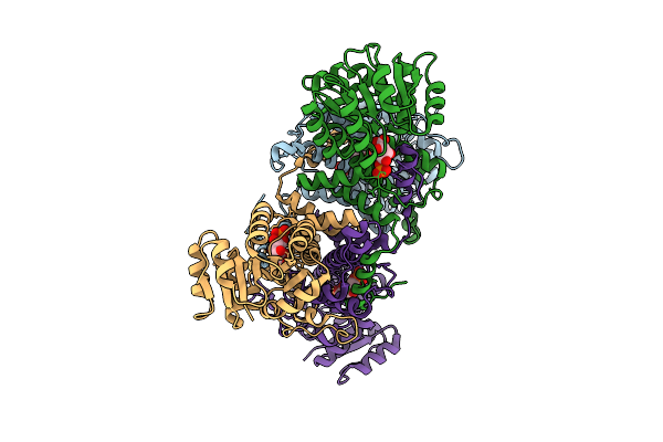 |
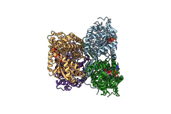
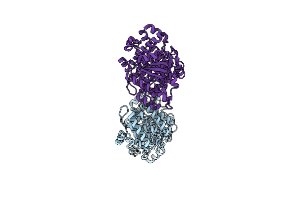
Crystal Structure Of Tetrameric 6-Phosphogluconate Dehydrogenase From Gluconobacter Oxydans In Complex With 6-Phosphogluconate
Organism: Gluconobacter oxydans
Method: X-RAY DIFFRACTION Resolution:2.00 Å Release Date: 2026-03-18 Classification: OXIDOREDUCTASE Ligands: 6PG |
Organism: Gluconobacter oxydans
Method: X-RAY DIFFRACTION
Release Date: 2026-03-18
Ligands: 6PG
 |
C-Terminally Truncated Dextran Dextrinase Bound With Acarbose
Organism: Gluconobacter oxydans
Method: X-RAY DIFFRACTION Resolution:2.50 Å Release Date: 2025-09-03 Classification: TRANSFERASE Ligands: EDO, CIT |
Organism: Gluconobacter oxydans
Method: X-RAY DIFFRACTION
Release Date: 2025-09-03
Ligands: EDO, CIT
 |
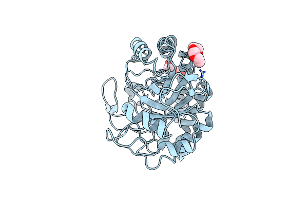
Gluer Mutant - W66F F269Y Q293T F68Y T36E P263L
Organism: Gluconobacter oxydans
Method: X-RAY DIFFRACTION Resolution:1.47 Å Release Date: 2024-03-13 Classification: OXIDOREDUCTASE Ligands: ACT, GOL, FMN |
Organism: Gluconobacter oxydans
Method: X-RAY DIFFRACTION
Release Date: 2024-03-13
Ligands: ACT, GOL, FMN
 |
1,3 L,D-Transpeptidase From Gluconobacter Oxydans
Organism: Gluconobacter oxydans
Method: X-RAY DIFFRACTION Resolution:1.73 Å Release Date: 2023-11-08 Classification: TRANSFERASE Ligands: CL, NA |
Organism: Gluconobacter oxydans
Method: X-RAY DIFFRACTION
Release Date: 2023-11-08
Ligands: CL, NA
 |
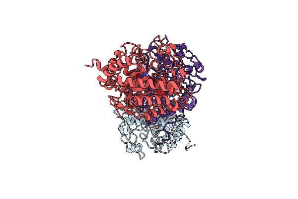
Cryo-Em Structure Of Membrane-Bound Alcohol Dehydrogenase From Gluconobacter Oxydans
Organism: Gluconobacter oxydans 621h, Gluconobacter oxydans
Method: ELECTRON MICROSCOPY Release Date: 2023-08-02 Classification: OXIDOREDUCTASE Ligands: HEC, PQQ, CA, U10 |
Organism: Gluconobacter oxydans 621h, Gluconobacter oxydans
Method: ELECTRON MICROSCOPY
Release Date: 2023-08-02
Ligands: HEC, PQQ, CA, U10
 |
Cryo-Em Structure Of Membrane-Bound Aldehyde Dehydrogenase From Gluconobacter Oxydans
Organism: Gluconobacter oxydans
Method: ELECTRON MICROSCOPY Release Date: 2023-08-02 Classification: OXIDOREDUCTASE Ligands: HEC, U10, FES, PCD |
Organism: Gluconobacter oxydans
Method: ELECTRON MICROSCOPY
Release Date: 2023-08-02
Ligands: HEC, U10, FES, PCD
 |
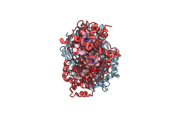
Gluconobacter Ene-Reductase (Gluer) Mutant - Pager
Organism: Gluconobacter oxydans
Method: X-RAY DIFFRACTION Resolution:1.50 Å Release Date: 2023-06-28 Classification: OXIDOREDUCTASE Ligands: FMN |
Organism: Gluconobacter oxydans
Method: X-RAY DIFFRACTION
Release Date: 2023-06-28
Ligands: FMN
 |
The C296A Mutant Of L-Sorbosone Dehydrogenase (Sndh) From Gluconobacter Oxydans Wsh-004
Organism: Gluconobacter oxydans
Method: X-RAY DIFFRACTION Resolution:2.22 Å Release Date: 2023-03-01 Classification: OXIDOREDUCTASE Ligands: NAP |
Organism: Gluconobacter oxydans
Method: X-RAY DIFFRACTION
Release Date: 2023-03-01
Ligands: NAP
 |
The Crystal Structure Of The Reduced Form Of Gluconobacter Oxydans Wsh-004 Sndh
Organism: Gluconobacter oxydans
Method: X-RAY DIFFRACTION Resolution:2.99 Å Release Date: 2023-03-01 Classification: OXIDOREDUCTASE Ligands: NDP |
Organism: Gluconobacter oxydans
Method: X-RAY DIFFRACTION
Release Date: 2023-03-01
Ligands: NDP
 |
The Crystal Structure Of The Oxidized Form Of Gluconobacter Oxydans Wsh-004 Sndh
Organism: Gluconobacter oxydans
Method: X-RAY DIFFRACTION Resolution:2.50 Å Release Date: 2023-01-18 Classification: OXIDOREDUCTASE |
Organism: Gluconobacter oxydans
Method: X-RAY DIFFRACTION
Release Date: 2023-01-18
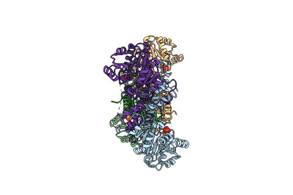 |
Crystal Structure Of The Tetrameric 6-Phosphogluconate Dehydrogenase From Gluconobacter Oxidans
Organism: Gluconobacter oxydans
Method: X-RAY DIFFRACTION Resolution:3.20 Å Release Date: 2020-12-02 Classification: OXIDOREDUCTASE Ligands: SO4 |
Organism: Gluconobacter oxydans
Method: X-RAY DIFFRACTION
Release Date: 2020-12-02
Ligands: SO4
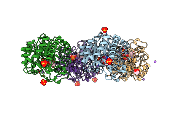 |
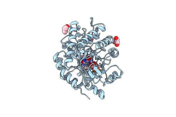
Gluconobacter Ene-Reductase (Gluer) Mutant - T36A
Organism: Gluconobacter oxydans
Method: X-RAY DIFFRACTION Resolution:1.16 Å Release Date: 2019-06-26 Classification: OXIDOREDUCTASE Ligands: FMN, ACT, SO4, NA, GOL |
Organism: Gluconobacter oxydans
Method: X-RAY DIFFRACTION
Release Date: 2019-06-26
Ligands: FMN, ACT, SO4, NA, GOL
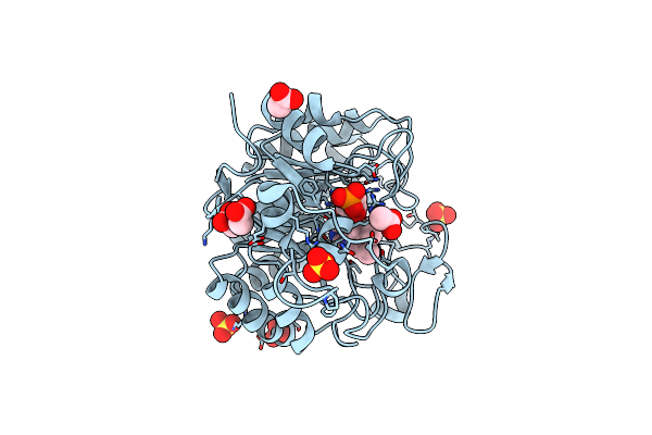 |
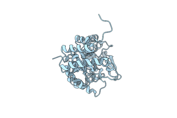
Gluconobacter Ene-Reductase (Gluer)
Organism: Gluconobacter oxydans
Method: X-RAY DIFFRACTION Resolution:1.80 Å Release Date: 2019-06-26 Classification: OXIDOREDUCTASE Ligands: FMN, SO4, ACT, NA, CL |
Organism: Gluconobacter oxydans
Method: X-RAY DIFFRACTION
Release Date: 2019-06-26
Ligands: FMN, SO4, ACT, NA, CL
 |
Crystal Structure Of D-Sorbitol Dehydrogenase In Substrate-Free Form
Organism: Gluconobacter oxydans
Method: X-RAY DIFFRACTION Resolution:1.95 Å Release Date: 2017-03-08 Classification: OXIDOREDUCTASE |
Organism: Gluconobacter oxydans
Method: X-RAY DIFFRACTION
Release Date: 2017-03-08
 |
Crystal Structure Of Gye (Old Yellow Enzyme)
Organism: Gluconobacter oxydans
Method: X-RAY DIFFRACTION Resolution:3.30 Å Release Date: 2014-09-24 Classification: OXIDOREDUCTASE ACTIVATOR Ligands: PGE, PEG, HG |
Organism: Gluconobacter oxydans
Method: X-RAY DIFFRACTION
Release Date: 2014-09-24
Ligands: PGE, PEG, HG
 |
Crystal Structure Of Gox2181
Organism: Gluconobacter oxydans
Method: X-RAY DIFFRACTION Resolution:1.80 Å Release Date: 2012-02-01 Classification: OXIDOREDUCTASE Ligands: MG, CD |
Organism: Gluconobacter oxydans
Method: X-RAY DIFFRACTION
Release Date: 2012-02-01
Ligands: MG, CD
 |
Structure Of The Glycerol Dehydrogenase Akr11B4 From Gluconobacter Oxydans
Organism: Gluconobacter oxydans
Method: X-RAY DIFFRACTION Resolution:2.00 Å Release Date: 2010-07-21 Classification: OXIDOREDUCTASE |
Organism: Gluconobacter oxydans
Method: X-RAY DIFFRACTION
Release Date: 2010-07-21
 |
Crystal Structure Of Nad+-Bound Xylitol Dehydrogenase
Organism: Gluconobacter oxydans
Method: X-RAY DIFFRACTION Resolution:1.90 Å Release Date: 2006-03-28 Classification: OXIDOREDUCTASE Ligands: NAD, MG |
Organism: Gluconobacter oxydans
Method: X-RAY DIFFRACTION
Release Date: 2006-03-28
Ligands: NAD, MG