

Search Count: 3,554
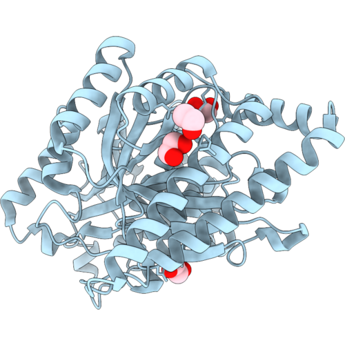 |
Mevalonate Kinase From Saccharomyces Cerevisiae
Organism: Saccharomyces cerevisiae
Method: X-RAY DIFFRACTION Resolution:2.15 Å Release Date: 2026-09-30 Classification: TRANSFERASE Ligands: EDO, GOL |
Organism: Saccharomyces cerevisiae
Method: X-RAY DIFFRACTION
Release Date: 2026-09-30
Ligands: EDO, GOL
 |
Mevalonate Kinase From Saccharomyces Cerevisiae With Isopentenyl Pyrophosphate (Ipp) Bound
Organism: Saccharomyces cerevisiae
Method: X-RAY DIFFRACTION Resolution:2.07 Å Release Date: 2026-09-30 Classification: TRANSFERASE Ligands: MG, IPE, EDO |
Organism: Saccharomyces cerevisiae
Method: X-RAY DIFFRACTION
Release Date: 2026-09-30
Ligands: MG, IPE, EDO
 |
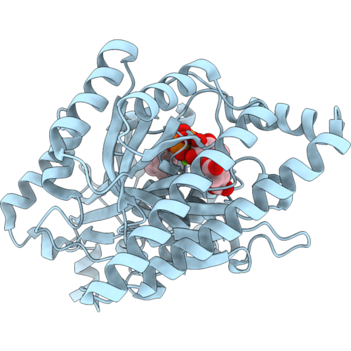
Mevalonate Kinase From Saccharomyces Cerevisiae With Dimethylallyl Pyrophosphate (Dmapp) Bound
Organism: Saccharomyces cerevisiae
Method: X-RAY DIFFRACTION Resolution:1.93 Å Release Date: 2026-09-30 Classification: TRANSFERASE Ligands: MG, DMA, TAR, EDO |
Organism: Saccharomyces cerevisiae
Method: X-RAY DIFFRACTION
Release Date: 2026-09-30
Ligands: MG, DMA, TAR, EDO
 |
Mevalonate Kinase From Saccharomyces Cerevisiae With Geranyl Pyrophosphate (Gpp) Bound
Organism: Saccharomyces cerevisiae
Method: X-RAY DIFFRACTION Resolution:2.05 Å Release Date: 2026-09-30 Classification: TRANSFERASE Ligands: MG, GPP, TAR, BGC |
Organism: Saccharomyces cerevisiae
Method: X-RAY DIFFRACTION
Release Date: 2026-09-30
Ligands: MG, GPP, TAR, BGC
 |
Mevalonate Kinase From Saccharomyces Cerevisiae With Farnesyl Pyrophosphate (Fpp) Bound
Organism: Saccharomyces cerevisiae
Method: X-RAY DIFFRACTION Resolution:2.09 Å Release Date: 2026-09-30 Classification: TRANSFERASE Ligands: FPP, MG, TAR |
Organism: Saccharomyces cerevisiae
Method: X-RAY DIFFRACTION
Release Date: 2026-09-30
Ligands: FPP, MG, TAR
 |
Mevalonate Kinase From Saccharomyces Cerevisiae With Geranylgeranyl Pyrophosphate (Ggpp) Bound
Organism: Saccharomyces cerevisiae
Method: X-RAY DIFFRACTION Resolution:1.95 Å Release Date: 2026-09-30 Classification: TRANSFERASE Ligands: MG, GRG, TAR, BGC, GOL |
Organism: Saccharomyces cerevisiae
Method: X-RAY DIFFRACTION
Release Date: 2026-09-30
Ligands: MG, GRG, TAR, BGC, GOL
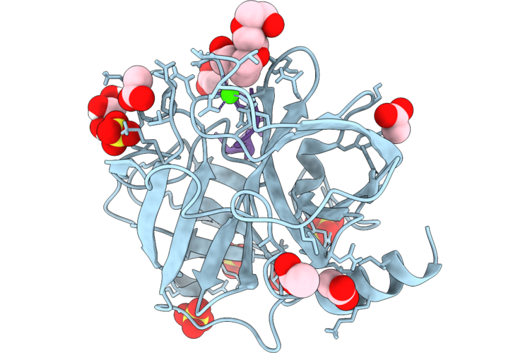 |
Sfti-1 Containing L-2-Thiaproline At Residue 8 In Complex With Trypsin
Organism: Helianthus annuus, Bos taurus
Method: X-RAY DIFFRACTION Resolution:1.47 Å Release Date: 2026-09-23 Classification: HYDROLASE/HYDROLASE INHIBITOR Ligands: ACT, CA, SO4, PG4, GOL |
Organism: Helianthus annuus, Bos taurus
Method: X-RAY DIFFRACTION
Release Date: 2026-09-23
Ligands: ACT, CA, SO4, PG4, GOL
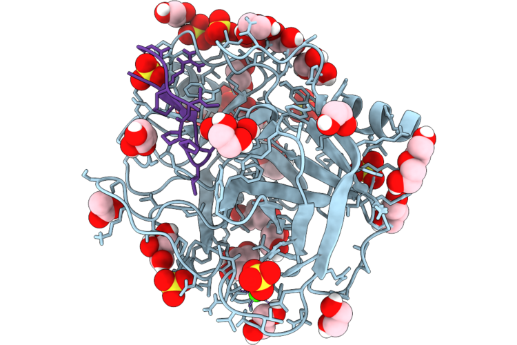 |
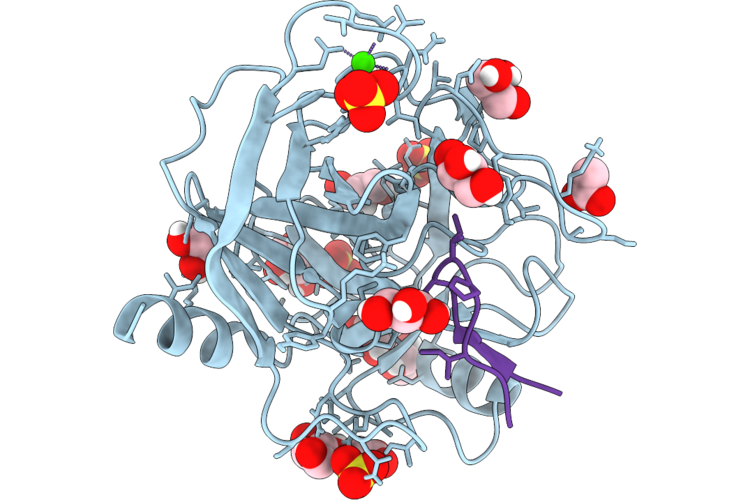
Sfti-1 Containing L-2-Thiaproline At Residue 9 In Complex With Trypsin
Organism: Helianthus annuus, Bos taurus
Method: X-RAY DIFFRACTION Resolution:1.22 Å Release Date: 2026-09-23 Classification: HYDROLASE/HYDROLASE INHIBITOR Ligands: CA, GOL, ACT, EDO, SO4, PEG |
Organism: Helianthus annuus, Bos taurus
Method: X-RAY DIFFRACTION
Release Date: 2026-09-23
Ligands: CA, GOL, ACT, EDO, SO4, PEG
 |
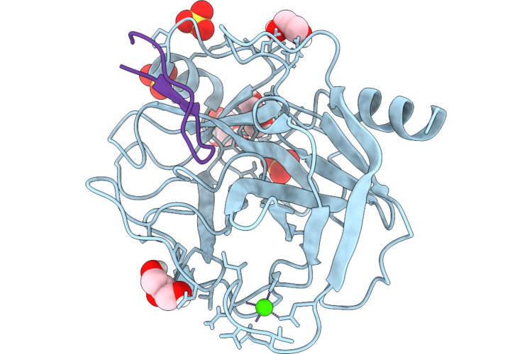
Sfti-1 Containing L-2-Thiaproline At Residue 13 In Complex With Trypsin
Organism: Helianthus annuus, Bos taurus
Method: X-RAY DIFFRACTION Resolution:1.13 Å Release Date: 2026-09-23 Classification: HYDROLASE/HYDROLASE INHIBITOR Ligands: SO4, CA, GOL, EDO, ACT |
Organism: Helianthus annuus, Bos taurus
Method: X-RAY DIFFRACTION
Release Date: 2026-09-23
Ligands: SO4, CA, GOL, EDO, ACT
 |
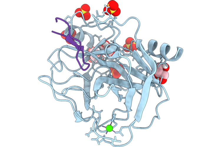
Sfti-1 Containing L-2-Selenaproline At Residue 8 In Complex With Trypsin
Organism: Helianthus annuus, Bos taurus
Method: X-RAY DIFFRACTION Resolution:1.17 Å Release Date: 2026-09-23 Classification: HYDROLASE/HYDROLASE INHIBITOR Ligands: CA, EDO, GOL, SO4 |
Organism: Helianthus annuus, Bos taurus
Method: X-RAY DIFFRACTION
Release Date: 2026-09-23
Ligands: CA, EDO, GOL, SO4
 |
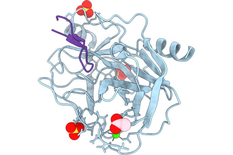
Sfti-1 Containing L-2-Selenaproline At Residue 9 In Complex With Trypsin
Organism: Helianthus annuus, Bos taurus
Method: X-RAY DIFFRACTION Resolution:1.45 Å Release Date: 2026-09-23 Classification: HYDROLASE/HYDROLASE INHIBITOR Ligands: CA, SO4, PO4, GOL, EDO |
Organism: Helianthus annuus, Bos taurus
Method: X-RAY DIFFRACTION
Release Date: 2026-09-23
Ligands: CA, SO4, PO4, GOL, EDO
 |
Sfti-1 Containing L-2-Selenaproline At Residue 13 In Complex With Trypsin
Organism: Helianthus annuus, Bos taurus
Method: X-RAY DIFFRACTION Resolution:1.90 Å Release Date: 2026-09-23 Classification: HYDROLASE/HYDROLASE INHIBITOR Ligands: CA, SO4, ACT |
Organism: Helianthus annuus, Bos taurus
Method: X-RAY DIFFRACTION
Release Date: 2026-09-23
Ligands: CA, SO4, ACT
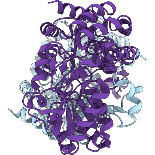 |
Crystal Structure Of Bovine Aldh3A1 H290L Mutant - Apo Form
Organism: Bos taurus
Method: X-RAY DIFFRACTION Resolution:1.40 Å Release Date: 2026-09-02 Classification: OXIDOREDUCTASE |
Organism: Bos taurus
Method: X-RAY DIFFRACTION
Release Date: 2026-09-02
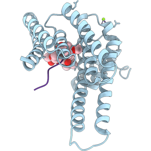 |
Ternary Structure Of 14-3-3Sigma, Interaction Cassette Phosphopeptide, And Fusicoccin-A
Organism: Homo sapiens, Synthetic construct
Method: X-RAY DIFFRACTION Resolution:1.40 Å Release Date: 2026-09-02 Classification: PEPTIDE BINDING PROTEIN Ligands: MG, CL, SIT |
Organism: Homo sapiens, Synthetic construct
Method: X-RAY DIFFRACTION
Release Date: 2026-09-02
Ligands: MG, CL, SIT
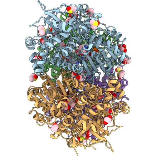 |
Crystal Structure Of Human Dhx8 In Complex With Compound 1 And Adp
Organism: Homo sapiens
Method: X-RAY DIFFRACTION Resolution:2.65 Å Release Date: 2026-09-02 Classification: RNA BINDING PROTEIN Ligands: ADP, MG, A1J1C, DMS, EDO |
Organism: Homo sapiens
Method: X-RAY DIFFRACTION
Release Date: 2026-09-02
Ligands: ADP, MG, A1J1C, DMS, EDO
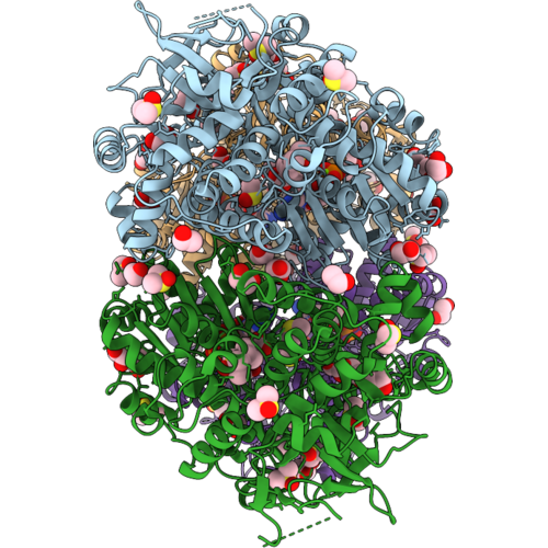 |
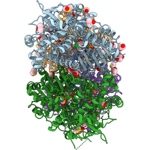
Crystal Structure Of Human Dhx8 In Complex With Compound 4 And Adp
Organism: Homo sapiens
Method: X-RAY DIFFRACTION Resolution:2.55 Å Release Date: 2026-09-02 Classification: RNA BINDING PROTEIN Ligands: ADP, MG, DMS, IBP, EDO, IZP, CL |
Organism: Homo sapiens
Method: X-RAY DIFFRACTION
Release Date: 2026-09-02
Ligands: ADP, MG, DMS, IBP, EDO, IZP, CL
 |
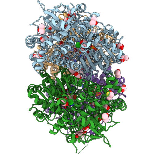
Crystal Structure Of Human Dhx8 In Complex With Compound 5 And Adp
Organism: Homo sapiens
Method: X-RAY DIFFRACTION Resolution:2.64 Å Release Date: 2026-09-02 Classification: RNA BINDING PROTEIN Ligands: ADP, MG, DMS, EDO, A1J1D |
Organism: Homo sapiens
Method: X-RAY DIFFRACTION
Release Date: 2026-09-02
Ligands: ADP, MG, DMS, EDO, A1J1D
 |
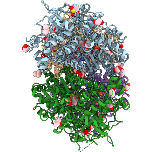
Crystal Structure Of Human Dhx8 In Complex With Compound 6 And Adp
Organism: Homo sapiens
Method: X-RAY DIFFRACTION Resolution:2.69 Å Release Date: 2026-09-02 Classification: RNA BINDING PROTEIN Ligands: ADP, MG, DMS, EDO, A1J1E |
Organism: Homo sapiens
Method: X-RAY DIFFRACTION
Release Date: 2026-09-02
Ligands: ADP, MG, DMS, EDO, A1J1E
 |
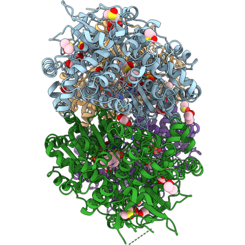
Crystal Structure Of Human Dhx8 In Complex With Compound 21 And Adp
Organism: Homo sapiens
Method: X-RAY DIFFRACTION Resolution:2.85 Å Release Date: 2026-09-02 Classification: RNA BINDING PROTEIN Ligands: ADP, MG, DMS, EDO, A1J1F |
Organism: Homo sapiens
Method: X-RAY DIFFRACTION
Release Date: 2026-09-02
Ligands: ADP, MG, DMS, EDO, A1J1F
 |
Crystal Structure Of Human Dhx8 In Complex With Compound 24 And Adp
Organism: Homo sapiens
Method: X-RAY DIFFRACTION Resolution:2.97 Å Release Date: 2026-09-02 Classification: RNA BINDING PROTEIN Ligands: ADP, MG, DMS, EDO, A1J1G |
Organism: Homo sapiens
Method: X-RAY DIFFRACTION
Release Date: 2026-09-02
Ligands: ADP, MG, DMS, EDO, A1J1G