
Search Count: 8,482
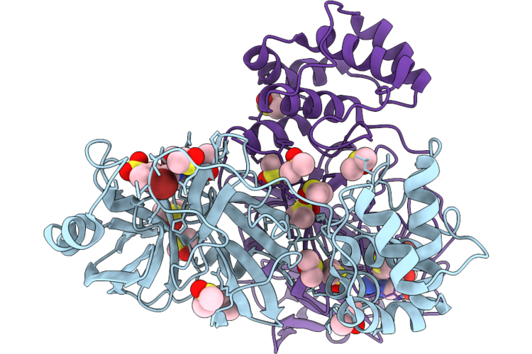 |
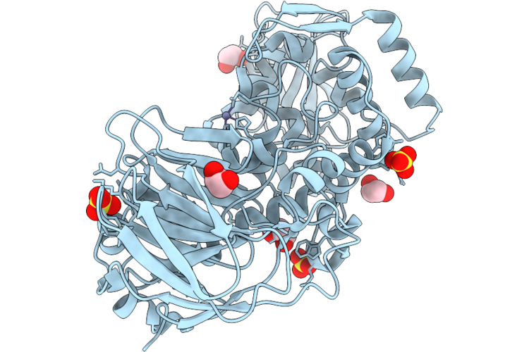
Pandda Deposition -- Crystal Structure Of Sars-Cov-2 Main Protease Covalently Bound To Kl_C172
Organism: Severe acute respiratory syndrome coronavirus 2
Method: X-RAY DIFFRACTION Resolution:1.93 Å Release Date: 2026-06-24 Classification: HYDROLASE Ligands: DMS, CL, A1J3E |
 |
Organism: Hiv-1 group m
Method: ELECTRON MICROSCOPY Resolution:3.75 Å Release Date: 2026-06-24 Classification: VIRAL PROTEIN Ligands: IHP |
 |
Organism: Hiv-1 group m
Method: ELECTRON MICROSCOPY Resolution:3.47 Å Release Date: 2026-06-24 Classification: ANTIMICROBIAL PROTEIN Ligands: IHP |
 |
Organism: Homo sapiens, Synthetic construct, Hiv-1 group m
Method: ELECTRON MICROSCOPY Resolution:4.64 Å Release Date: 2026-06-24 Classification: ANTIVIRAL PROTEIN |
 |
Organism: Hiv-1 group m
Method: ELECTRON MICROSCOPY Resolution:4.10 Å Release Date: 2026-06-24 Classification: VIRAL PROTEIN |
 |
Structure Of The 4-Chrd Domain Of Human Chordin In Complex With A Heparin Oligosaccharide
Organism: Homo sapiens
Method: X-RAY DIFFRACTION Resolution:2.92 Å Release Date: 2026-06-24 Classification: UNKNOWN FUNCTION Ligands: BMA, NAG, CA, SO4 |
 |
Organism: Homo sapiens
Method: ELECTRON MICROSCOPY Release Date: 2026-06-24 Classification: SIGNALING PROTEIN/IMMUNE SYSTEM |
 |
C1 Symmetry Cryoem Structure Of The Soluble-Wraped Membranous Portion Of Mspa (Mycobacterium Smegmatis Porin), Dimerized Along The Native Interface.
Organism: Mycolicibacterium smegmatis mc2 155
Method: ELECTRON MICROSCOPY Resolution:3.34 Å Release Date: 2026-06-24 Classification: DE NOVO PROTEIN |
 |
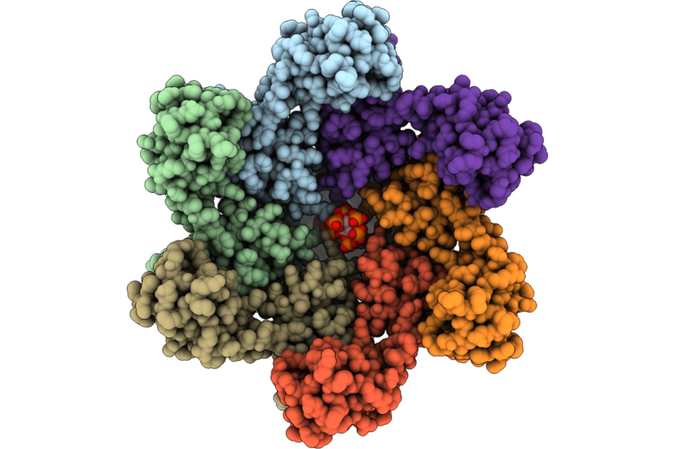
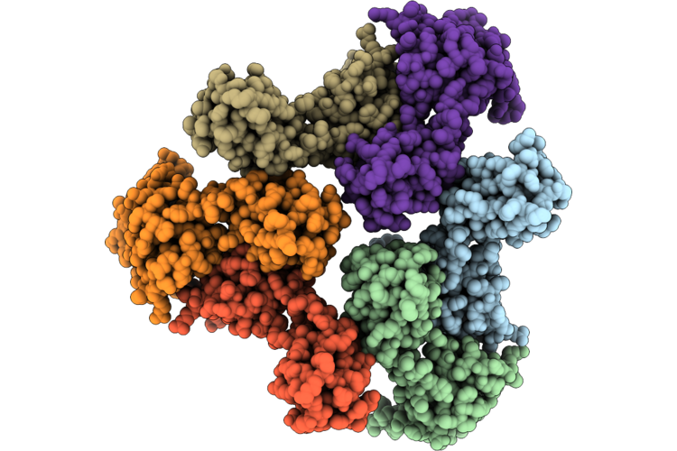
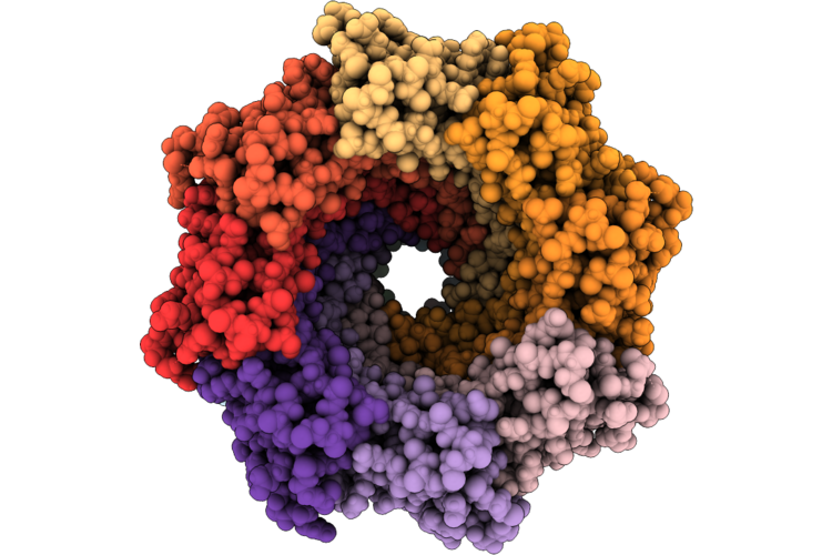
Cryo-Electron Microscopy Structure Of Pfripr Bound To Monoclonal Antibodies Rp.047, Rp.057 And Rp.035
Organism: Mus musculus, Plasmodium falciparum 3d7
Method: ELECTRON MICROSCOPY Resolution:3.35 Å Release Date: 2026-06-24 Classification: IMMUNE SYSTEM |
 |
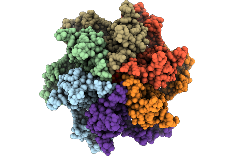
Cryo-Electron Microscopy Structure Of Pfripr Bound To Monoclonal Antibodies Rp.093, Rp.073 And Rp.063
Organism: Mus musculus, Plasmodium falciparum 3d7
Method: ELECTRON MICROSCOPY Resolution:2.97 Å Release Date: 2026-06-24 Classification: IMMUNE SYSTEM |
 |
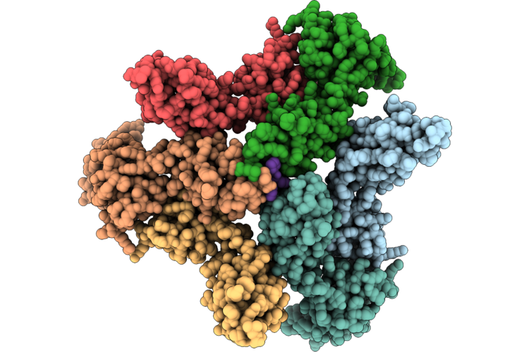
Cryo-Electron Microscopy Structure Of Pfripr Bound To Monoclonal Antibodies Rp.092 And Rp.052
Organism: Plasmodium falciparum 3d7, Mus musculus
Method: ELECTRON MICROSCOPY Resolution:4.01 Å Release Date: 2026-06-24 Classification: IMMUNE SYSTEM |
 |
Organism: Homo sapiens
Method: ELECTRON MICROSCOPY Release Date: 2026-06-24 Classification: BLOOD CLOTTING Ligands: NAG |
 |
Organism: Homo sapiens
Method: ELECTRON MICROSCOPY Release Date: 2026-06-24 Classification: BLOOD CLOTTING Ligands: NAG |
 |
Organism: Bacteroides caccae atcc 43185
Method: X-RAY DIFFRACTION Resolution:1.80 Å Release Date: 2026-06-17 Classification: HYDROLASE Ligands: ZN, EDO |
 |
Organism: Bacteroides caccae atcc 43185
Method: X-RAY DIFFRACTION Resolution:1.80 Å Release Date: 2026-06-17 Classification: HYDROLASE Ligands: ZN, EDO, MLI, CA, SIN |
 |
Organism: Bacteroides caccae atcc 43185
Method: X-RAY DIFFRACTION Resolution:2.10 Å Release Date: 2026-06-17 Classification: HYDROLASE Ligands: ZN, EDO, BTB |
 |
Organism: Bacteroides caccae atcc 43185
Method: X-RAY DIFFRACTION Resolution:1.55 Å Release Date: 2026-06-17 Classification: HYDROLASE Ligands: ZN, SO4, EDO |
 |
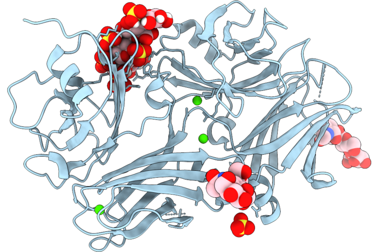
The Structure Of O-Glycopeptidase Bcm60C (E665A Mutant) From Bacteroides Caccae In Complex With A Core 2 Glycan
Organism: Bacteroides caccae atcc 43185
Method: X-RAY DIFFRACTION Resolution:2.15 Å Release Date: 2026-06-17 Classification: HYDROLASE Ligands: THR, ZN, SO4, EDO |
 |
The Structure Of O-Glycopeptidase Bcm60K From Bacteroides Caccae In Complex With A Core 2 Glycan
Organism: Bacteroides caccae atcc 43185, Homo sapiens
Method: X-RAY DIFFRACTION Resolution:2.19 Å Release Date: 2026-06-17 Classification: HYDROLASE Ligands: ZN, A2G |
 |
The Structure Of O-Glycopeptidase Bcm60K From Bacteroides Caccae In Complex With A Core 2 Glycan
Organism: Bacteroides caccae atcc 43185
Method: X-RAY DIFFRACTION Resolution:2.49 Å Release Date: 2026-06-17 Classification: HYDROLASE Ligands: THR, ZN |