

Search Count: 5,098
 |
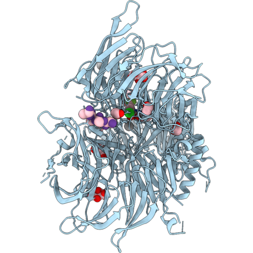
Txgh116 Complex With A Beta-D-Arabinofuranose-Configured Cyclitol Aziridine
Organism: Thermoanaerobacterium xylanolyticum lx-11
Method: X-RAY DIFFRACTION Resolution:1.75 Å Release Date: 2026-09-30 Classification: HYDROLASE Ligands: EDO, A1J9P, SO4, CA |
Organism: Thermoanaerobacterium xylanolyticum lx-11
Method: X-RAY DIFFRACTION
Release Date: 2026-09-30
Ligands: EDO, A1J9P, SO4, CA
 |
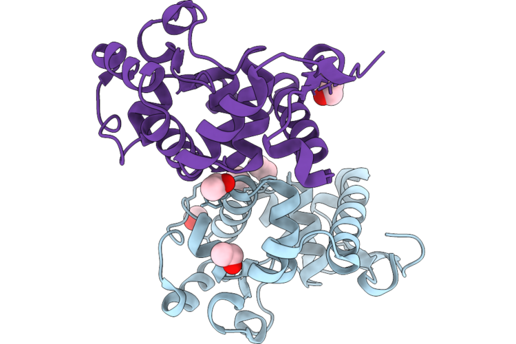
Crystal Structure Of Ddb1 In Complex With Xs445479
Organism: Homo sapiens
Method: X-RAY DIFFRACTION Resolution:2.10 Å Release Date: 2026-09-30 Classification: LIGASE Ligands: EDO, TLA, A1DPN, UNX |
Organism: Homo sapiens
Method: X-RAY DIFFRACTION
Release Date: 2026-09-30
Ligands: EDO, TLA, A1DPN, UNX
 |
Crystal Structure Of The Gh134 Mannanase From Aspergillus Nidulans
Organism: Aspergillus nidulans fgsc a4
Method: X-RAY DIFFRACTION Resolution:1.75 Å Release Date: 2026-09-23 Classification: CARBOHYDRATE Ligands: EOH, POL |
Organism: Aspergillus nidulans fgsc a4
Method: X-RAY DIFFRACTION
Release Date: 2026-09-23
Ligands: EOH, POL
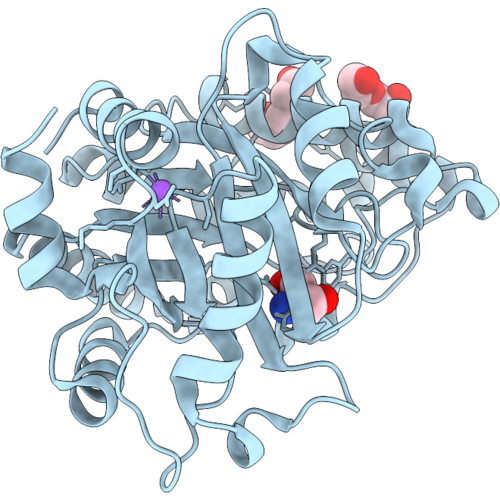 |
Crystal Structure Of A Gh26 Enzyme (Eigh26A)
Organism: Metagenome
Method: X-RAY DIFFRACTION Resolution:1.08 Å Release Date: 2026-09-09 Classification: HYDROLASE Ligands: TRS, PEG, PGE, NA |
Organism: Metagenome
Method: X-RAY DIFFRACTION
Release Date: 2026-09-09
Ligands: TRS, PEG, PGE, NA
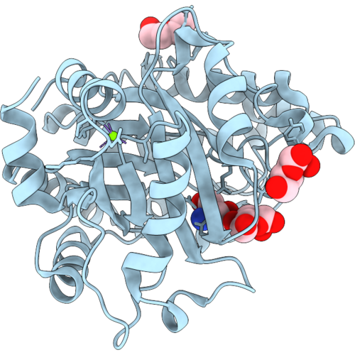 |
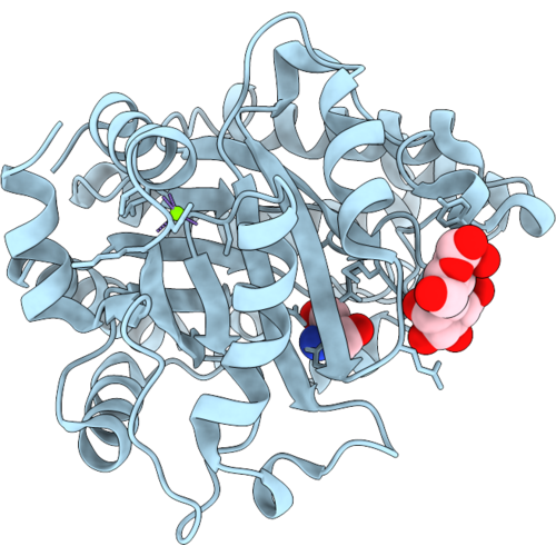
Crystal Structure Of A Gh26 Enzyme (Eigh26A) In Complex With Mannose
Organism: Metagenome
Method: X-RAY DIFFRACTION Resolution:1.12 Å Release Date: 2026-09-09 Classification: HYDROLASE Ligands: BMA, TRS, PEG, MG |
Organism: Metagenome
Method: X-RAY DIFFRACTION
Release Date: 2026-09-09
Ligands: BMA, TRS, PEG, MG
 |
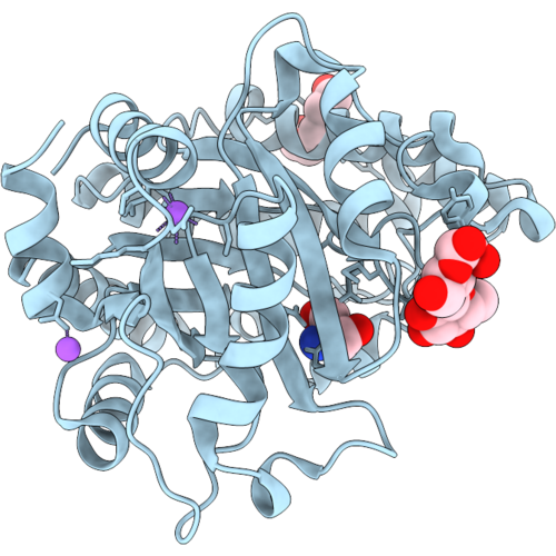
Crystal Structure Of A Gh26 Enzyme (Eigh26A) In Complex With Mannobiose
Organism: Metagenome
Method: X-RAY DIFFRACTION Resolution:1.07 Å Release Date: 2026-09-09 Classification: HYDROLASE Ligands: TRS, MG |
Organism: Metagenome
Method: X-RAY DIFFRACTION
Release Date: 2026-09-09
Ligands: TRS, MG
 |
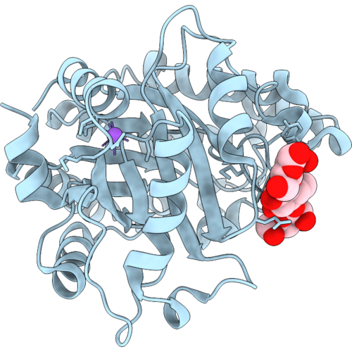
Crystal Structure Of A Gh26 Enzyme (Eigh26A) In Complex With Glucose-Beta-1,4-Mannose
Organism: Metagenome
Method: X-RAY DIFFRACTION Resolution:1.20 Å Release Date: 2026-09-09 Classification: HYDROLASE |
Organism: Metagenome
Method: X-RAY DIFFRACTION
Release Date: 2026-09-09
 |
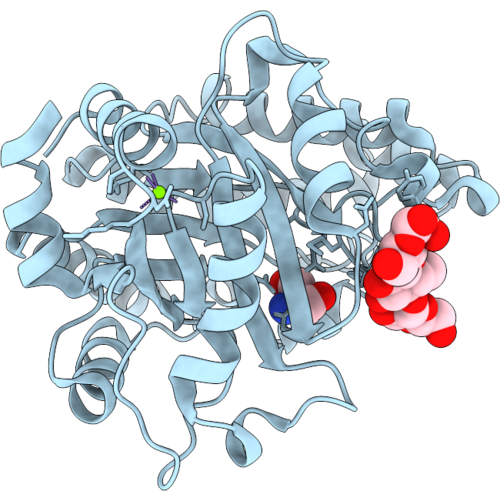
Crystal Structure Of A Gh26 Enzyme (Eigh26A) In Complex With Mannose-Beta-1,4-Glucose-Beta-1,4-Mannose
Organism: Metagenome
Method: X-RAY DIFFRACTION Resolution:1.60 Å Release Date: 2026-09-09 Classification: HYDROLASE |
Organism: Metagenome
Method: X-RAY DIFFRACTION
Release Date: 2026-09-09
 |
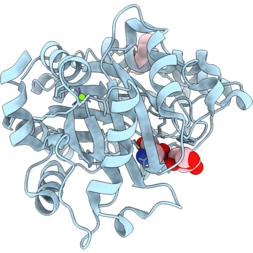
Crystal Structure Of A Gh26 Enzyme (Eigh26A) In Complex With Glucose-Beta-1,4-Glucose-Beta-1,4-Mannose
Organism: Metagenome
Method: X-RAY DIFFRACTION Resolution:1.18 Å Release Date: 2026-09-09 Classification: HYDROLASE Ligands: TRS, MG |
Organism: Metagenome
Method: X-RAY DIFFRACTION
Release Date: 2026-09-09
Ligands: TRS, MG
 |
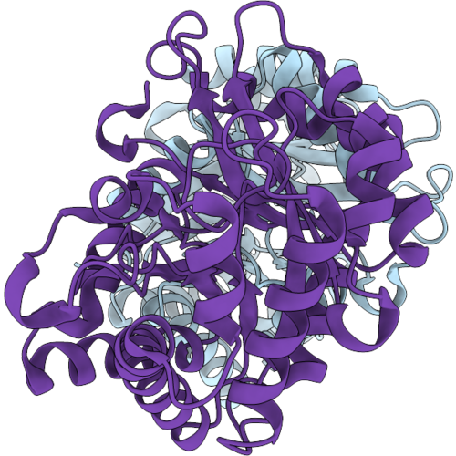
Crystal Structure Of A Gh26 Enzyme (Eigh26A) In Complex With Galactose
Organism: Metagenome
Method: X-RAY DIFFRACTION Resolution:1.53 Å Release Date: 2026-09-09 Classification: HYDROLASE Ligands: MG, PGE, GAL, TRS |
Organism: Metagenome
Method: X-RAY DIFFRACTION
Release Date: 2026-09-09
Ligands: MG, PGE, GAL, TRS
 |
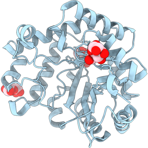
Crystal Structure Of A Gh26 Enzyme (Eigh26B)
Organism: Metagenome
Method: X-RAY DIFFRACTION Resolution:1.95 Å Release Date: 2026-09-09 Classification: HYDROLASE |
Organism: Metagenome
Method: X-RAY DIFFRACTION
Release Date: 2026-09-09
 |
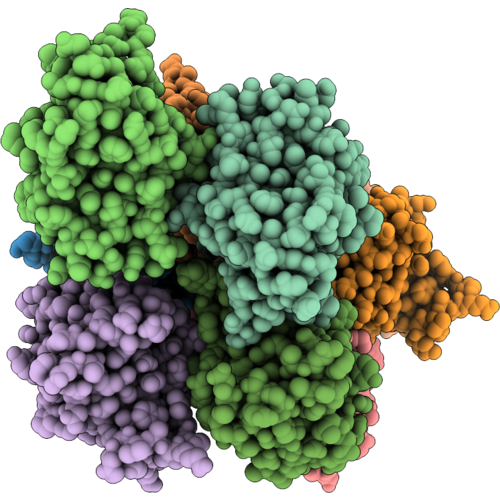
Crystal Structure Of A Gh26 Enzyme (Eigh26B) In Complex With Mannose
Organism: Metagenome
Method: X-RAY DIFFRACTION Resolution:1.71 Å Release Date: 2026-09-09 Classification: HYDROLASE Ligands: MAN, BMA |
Organism: Metagenome
Method: X-RAY DIFFRACTION
Release Date: 2026-09-09
Ligands: MAN, BMA
 |
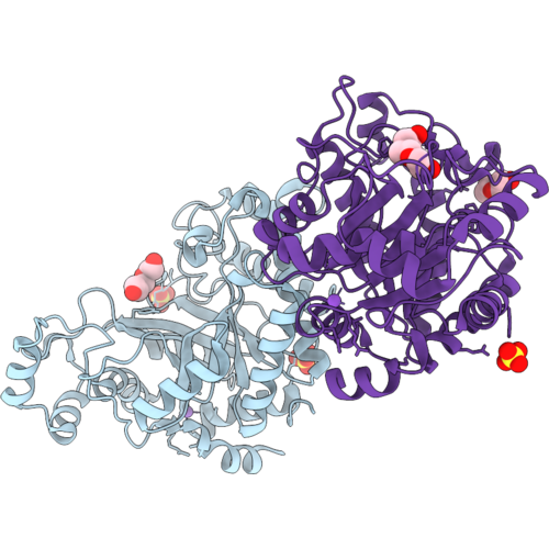
Crystal Structure Of A Beta-Domain (Beta-Dom_N1) From Gh26 (Eigh26B)
Organism: Metagenome
Method: X-RAY DIFFRACTION Resolution:2.30 Å Release Date: 2026-09-09 Classification: HYDROLASE |
Organism: Metagenome
Method: X-RAY DIFFRACTION
Release Date: 2026-09-09
 |
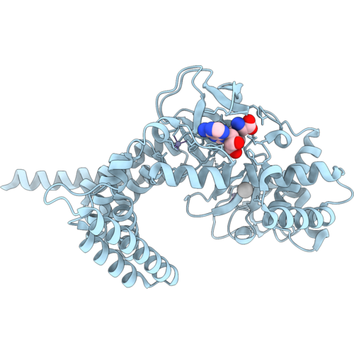
Crystal Structure Of A Gh26 Enzyme (Eigh26C)
Organism: Metagenome
Method: X-RAY DIFFRACTION Resolution:1.77 Å Release Date: 2026-09-09 Classification: HYDROLASE |
Organism: Metagenome
Method: X-RAY DIFFRACTION
Release Date: 2026-09-09
 |
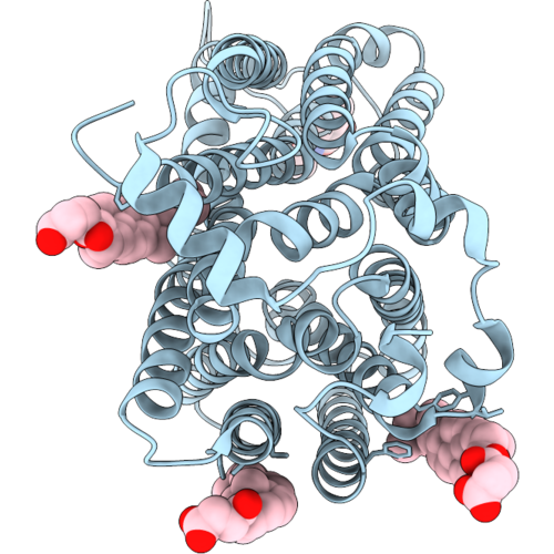
Crystal Structure Of Human Smyd1 In Complex With Sinefungin
Organism: Homo sapiens
Method: X-RAY DIFFRACTION Resolution:2.68 Å Release Date: 2026-09-02 Classification: TRANSFERASE Ligands: ZN, SFG, UNX |
Organism: Homo sapiens
Method: X-RAY DIFFRACTION
Release Date: 2026-09-02
Ligands: ZN, SFG, UNX
 |
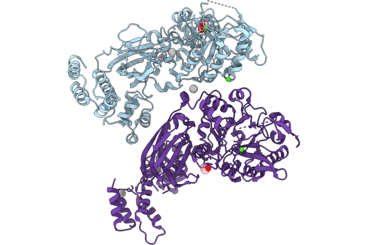
Cryo-Em Structure Of Vglut3 In Apo State
Organism: Mus musculus
Method: ELECTRON MICROSCOPY Release Date: 2026-09-02 Classification: TRANSPORT PROTEIN Ligands: NAG, Y01 |
Organism: Mus musculus
Method: ELECTRON MICROSCOPY
Release Date: 2026-09-02
Ligands: NAG, Y01
 |
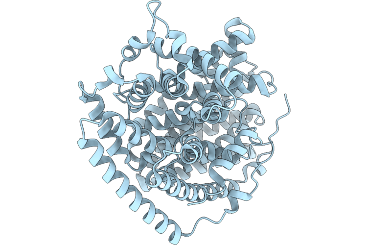
X-Ray Structure Of Chicken Plcz1 With Alternate Ef-Hand Conformation, Active Site Blocked By Phosphorylated Threonine In The Xy Linker
Organism: Gallus gallus
Method: X-RAY DIFFRACTION Resolution:1.99 Å Release Date: 2026-08-26 Classification: HYDROLASE Ligands: CL, CA, UNX, EDO |
Organism: Gallus gallus
Method: X-RAY DIFFRACTION
Release Date: 2026-08-26
Ligands: CL, CA, UNX, EDO
 |
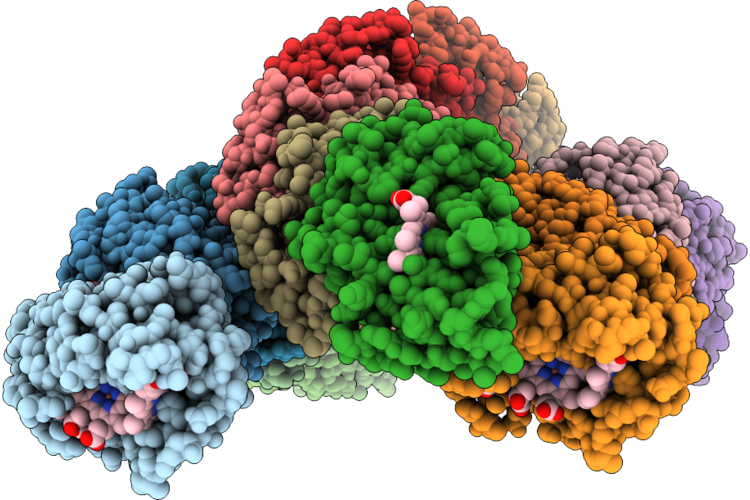
Drimenol Synthase From Persicaria Hydropiper
Organism: Persicaria hydropiper
Method: X-RAY DIFFRACTION Resolution:1.58 Å Release Date: 2026-08-26 Classification: BIOSYNTHETIC PROTEIN |
Organism: Persicaria hydropiper
Method: X-RAY DIFFRACTION
Release Date: 2026-08-26
 |
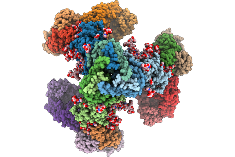
A Bundled Antiparallel Cytochrome Nanowire Produced By Desulfuromonas Soudanensis Wtl
Organism: Desulfuromonas soudanensis
Method: ELECTRON MICROSCOPY Resolution:3.11 Å Release Date: 2026-08-26 Classification: ELECTRON TRANSPORT Ligands: HEC, CA |
Organism: Desulfuromonas soudanensis
Method: ELECTRON MICROSCOPY
Release Date: 2026-08-26
Ligands: HEC, CA
 |
Composite Map Of Three Pairs Of Dimeric Vrc36 Fabs Bound To Hiv-1 Bg505.T332N Ds-Sosip.664 Env Trimer
Organism: Human immunodeficiency virus 1, Homo sapiens
Method: ELECTRON MICROSCOPY Resolution:3.59 Å Release Date: 2026-08-19 Classification: VIRAL PROTEIN/IMMUNE SYSTEM Ligands: NAG |
Organism: Human immunodeficiency virus 1, Homo sapiens
Method: ELECTRON MICROSCOPY
Release Date: 2026-08-19
Ligands: NAG