

Search Count: 3,277
 |
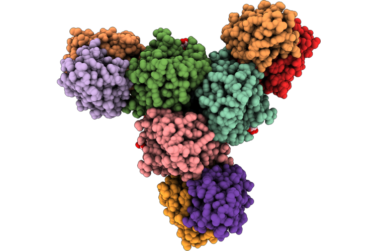
Structure Of Mink-Derived Hku5 Rbd In Complex With Ace2 Of N. Vison
Organism: Neogale vison, Pipistrellus bat coronavirus hku5
Method: ELECTRON MICROSCOPY Resolution:3.30 Å Release Date: 2026-09-30 Classification: VIRAL PROTEIN Ligands: NAG |
Organism: Neogale vison, Pipistrellus bat coronavirus hku5
Method: ELECTRON MICROSCOPY
Release Date: 2026-09-30
Ligands: NAG
 |
Cryoem Structure Of Monoclonal Fab 047-09 1A02 Binding The Lateral Patch Of Influenza Virus H1 Ha (A/California/04/2009 H1N1)
Organism: Influenza a virus (a/california/04/2009(h1n1)), Homo sapiens
Method: ELECTRON MICROSCOPY Release Date: 2026-09-23 Classification: IMMUNE SYSTEM/Viral Protein Ligands: NAG |
Organism: Influenza a virus (a/california/04/2009(h1n1)), Homo sapiens
Method: ELECTRON MICROSCOPY
Release Date: 2026-09-23
Ligands: NAG
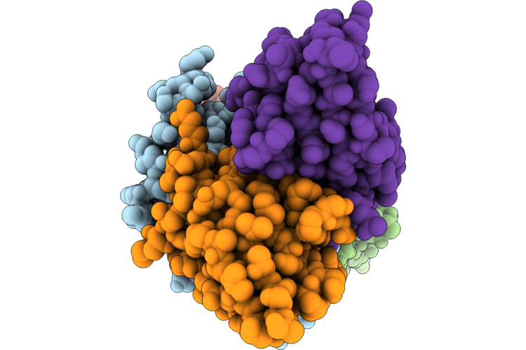 |
Hb3Var03 Cidra1.4 In Complex With B57 And C50 Fabs
Organism: Homo sapiens, Plasmodium falciparum hb3
Method: ELECTRON MICROSCOPY Resolution:3.40 Å Release Date: 2026-09-23 Classification: IMMUNE SYSTEM |
Organism: Homo sapiens, Plasmodium falciparum hb3
Method: ELECTRON MICROSCOPY
Release Date: 2026-09-23
 |
Structure Of Human Eepd1 Nuclease Domain
Organism: Homo sapiens
Method: X-RAY DIFFRACTION Resolution:3.20 Å Release Date: 2026-09-23 Classification: HYDROLASE |
Organism: Homo sapiens
Method: X-RAY DIFFRACTION
Release Date: 2026-09-23
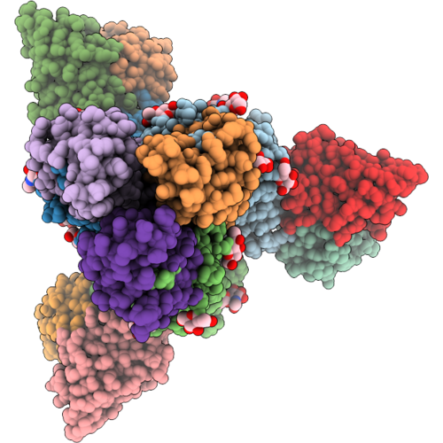 |
Cryoem Structure Of Monoclonal Fab 047-09M 2F03 Binding The Lateral Patch Of Influenza Virus H1 Ha (A/California/04/2009)
Organism: Homo sapiens
Method: ELECTRON MICROSCOPY Release Date: 2026-09-16 Classification: IMMUNE SYSTEM Ligands: NAG |
Organism: Homo sapiens
Method: ELECTRON MICROSCOPY
Release Date: 2026-09-16
Ligands: NAG
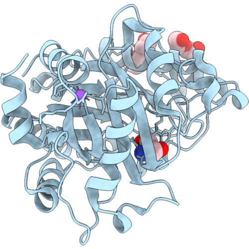 |
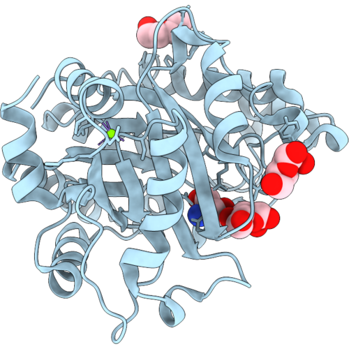
Crystal Structure Of A Gh26 Enzyme (Eigh26A)
Organism: Metagenome
Method: X-RAY DIFFRACTION Resolution:1.08 Å Release Date: 2026-09-09 Classification: HYDROLASE Ligands: TRS, PEG, PGE, NA |
Organism: Metagenome
Method: X-RAY DIFFRACTION
Release Date: 2026-09-09
Ligands: TRS, PEG, PGE, NA
 |
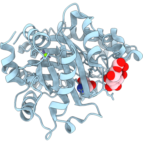
Crystal Structure Of A Gh26 Enzyme (Eigh26A) In Complex With Mannose
Organism: Metagenome
Method: X-RAY DIFFRACTION Resolution:1.12 Å Release Date: 2026-09-09 Classification: HYDROLASE Ligands: BMA, TRS, PEG, MG |
Organism: Metagenome
Method: X-RAY DIFFRACTION
Release Date: 2026-09-09
Ligands: BMA, TRS, PEG, MG
 |
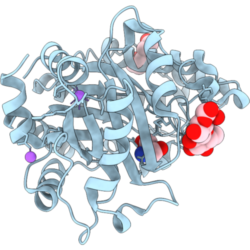
Crystal Structure Of A Gh26 Enzyme (Eigh26A) In Complex With Mannobiose
Organism: Metagenome
Method: X-RAY DIFFRACTION Resolution:1.07 Å Release Date: 2026-09-09 Classification: HYDROLASE Ligands: TRS, MG |
Organism: Metagenome
Method: X-RAY DIFFRACTION
Release Date: 2026-09-09
Ligands: TRS, MG
 |
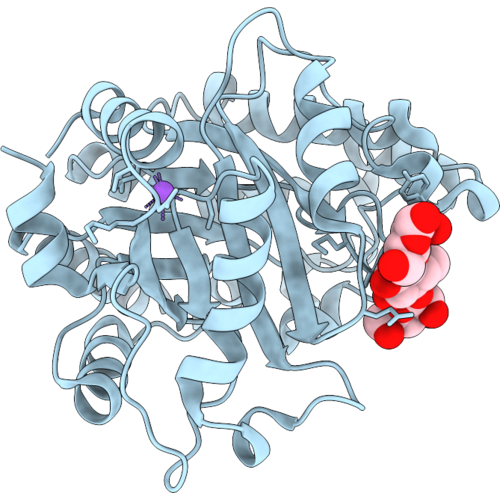
Crystal Structure Of A Gh26 Enzyme (Eigh26A) In Complex With Glucose-Beta-1,4-Mannose
Organism: Metagenome
Method: X-RAY DIFFRACTION Resolution:1.20 Å Release Date: 2026-09-09 Classification: HYDROLASE |
Organism: Metagenome
Method: X-RAY DIFFRACTION
Release Date: 2026-09-09
 |
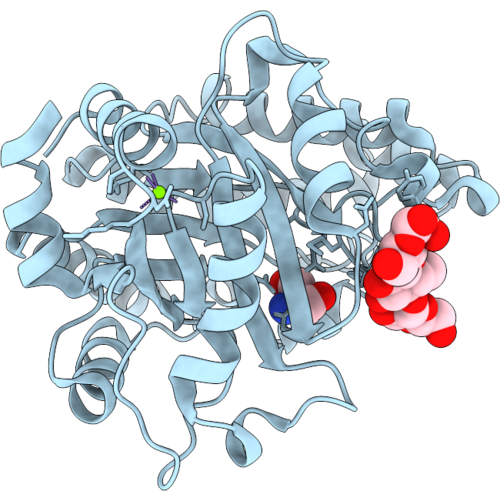
Crystal Structure Of A Gh26 Enzyme (Eigh26A) In Complex With Mannose-Beta-1,4-Glucose-Beta-1,4-Mannose
Organism: Metagenome
Method: X-RAY DIFFRACTION Resolution:1.60 Å Release Date: 2026-09-09 Classification: HYDROLASE |
Organism: Metagenome
Method: X-RAY DIFFRACTION
Release Date: 2026-09-09
 |
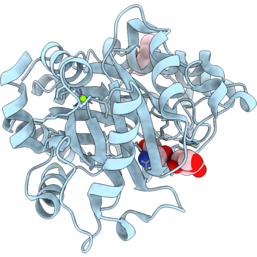
Crystal Structure Of A Gh26 Enzyme (Eigh26A) In Complex With Glucose-Beta-1,4-Glucose-Beta-1,4-Mannose
Organism: Metagenome
Method: X-RAY DIFFRACTION Resolution:1.18 Å Release Date: 2026-09-09 Classification: HYDROLASE Ligands: TRS, MG |
Organism: Metagenome
Method: X-RAY DIFFRACTION
Release Date: 2026-09-09
Ligands: TRS, MG
 |
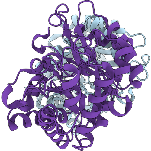
Crystal Structure Of A Gh26 Enzyme (Eigh26A) In Complex With Galactose
Organism: Metagenome
Method: X-RAY DIFFRACTION Resolution:1.53 Å Release Date: 2026-09-09 Classification: HYDROLASE Ligands: MG, PGE, GAL, TRS |
Organism: Metagenome
Method: X-RAY DIFFRACTION
Release Date: 2026-09-09
Ligands: MG, PGE, GAL, TRS
 |
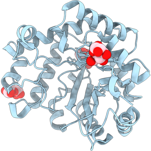
Crystal Structure Of A Gh26 Enzyme (Eigh26B)
Organism: Metagenome
Method: X-RAY DIFFRACTION Resolution:1.95 Å Release Date: 2026-09-09 Classification: HYDROLASE |
Organism: Metagenome
Method: X-RAY DIFFRACTION
Release Date: 2026-09-09
 |
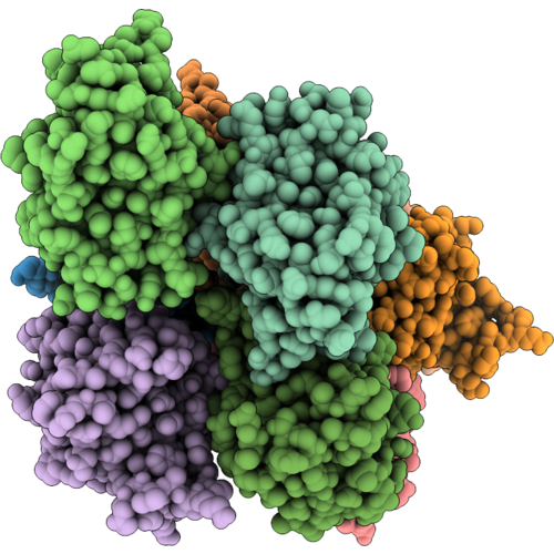
Crystal Structure Of A Gh26 Enzyme (Eigh26B) In Complex With Mannose
Organism: Metagenome
Method: X-RAY DIFFRACTION Resolution:1.71 Å Release Date: 2026-09-09 Classification: HYDROLASE Ligands: MAN, BMA |
Organism: Metagenome
Method: X-RAY DIFFRACTION
Release Date: 2026-09-09
Ligands: MAN, BMA
 |
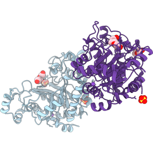
Crystal Structure Of A Beta-Domain (Beta-Dom_N1) From Gh26 (Eigh26B)
Organism: Metagenome
Method: X-RAY DIFFRACTION Resolution:2.30 Å Release Date: 2026-09-09 Classification: HYDROLASE |
Organism: Metagenome
Method: X-RAY DIFFRACTION
Release Date: 2026-09-09
 |
Crystal Structure Of A Gh26 Enzyme (Eigh26C)
Organism: Metagenome
Method: X-RAY DIFFRACTION Resolution:1.77 Å Release Date: 2026-09-09 Classification: HYDROLASE |
Organism: Metagenome
Method: X-RAY DIFFRACTION
Release Date: 2026-09-09
 |
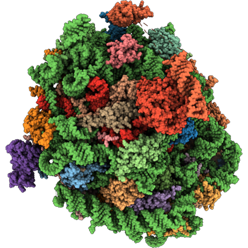
Cryo-Em Structure Of Donepi, A Deoxynivalenol 3-Epimerase From Nocardioides Sp. S5-5
Organism: Nocardioides sp. s5-5
Method: ELECTRON MICROSCOPY Resolution:2.60 Å Release Date: 2026-09-09 Classification: OXIDOREDUCTASE Ligands: NAP |
Organism: Nocardioides sp. s5-5
Method: ELECTRON MICROSCOPY
Release Date: 2026-09-09
Ligands: NAP
 |
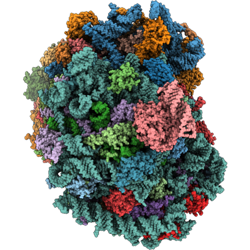
Rabbit 60S Ribosomal Subunit With Eef2 Domain Iv Closed
Organism: Oryctolagus cuniculus
Method: ELECTRON MICROSCOPY Release Date: 2026-09-02 Classification: RIBOSOME Ligands: ZN, GDP |
Organism: Oryctolagus cuniculus
Method: ELECTRON MICROSCOPY
Release Date: 2026-09-02
Ligands: ZN, GDP
 |
Rabbit 80S With Eef2,Ccdc124,And Larp1,40S-Head-Swiveled
Organism: Oryctolagus cuniculus
Method: ELECTRON MICROSCOPY Release Date: 2026-09-02 Classification: RIBOSOME Ligands: ZN, GDP |
Organism: Oryctolagus cuniculus
Method: ELECTRON MICROSCOPY
Release Date: 2026-09-02
Ligands: ZN, GDP
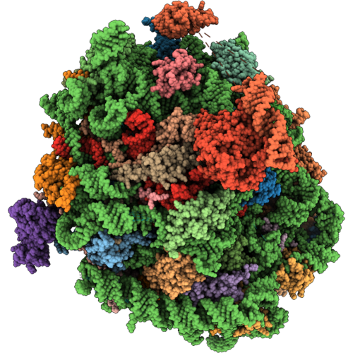 |
Rabbit 60S Ribosomal Subunit With Eef2 Domain Iv Open
Organism: Oryctolagus cuniculus
Method: ELECTRON MICROSCOPY Release Date: 2026-09-02 Classification: RIBOSOME Ligands: GDP, ZN |
Organism: Oryctolagus cuniculus
Method: ELECTRON MICROSCOPY
Release Date: 2026-09-02
Ligands: GDP, ZN