

9Z6X image
Deposition Date 2025-11-14
Release Date 2026-09-23
Last Version Date 2026-09-23
Entry Detail
PDB ID:
9Z6X
Keywords:
Title:
Structure of human EEPD1 nuclease domain
Biological Source:
Source Organism(s):
Homo sapiens (Taxon ID: 9606)
Expression System(s):
Method Details:
Experimental Method:
Resolution:
3.20 Å
R-Value Free:
0.28
R-Value Work:
0.25
R-Value Observed:
0.25
Space Group:
P 1 21 1
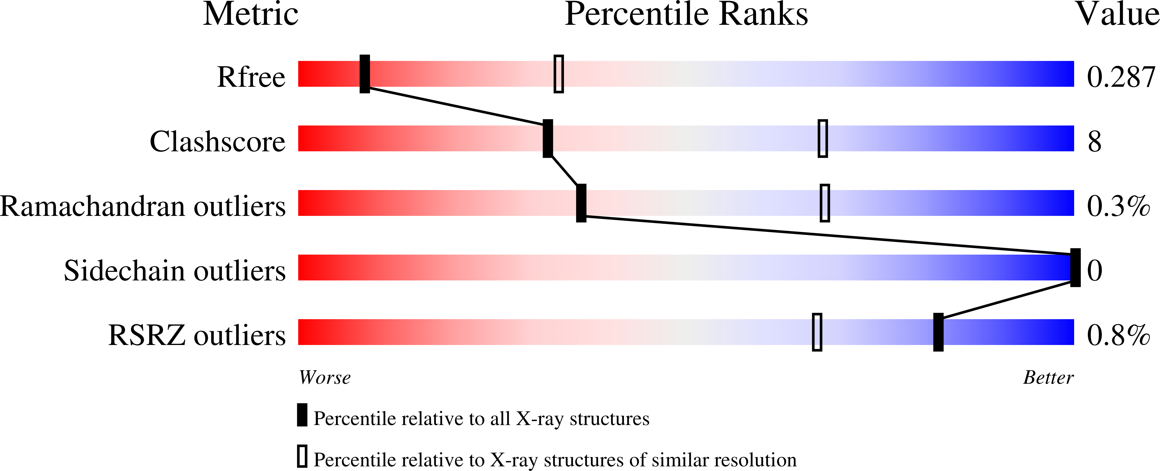
Macromolecular Entities
Polymer Type:polypeptide(L)
Molecule:Endonuclease/exonuclease/phos
Gene (Uniprot):EEPD1
Chain IDs:A, B (auth: C), C (auth: B), D
Chain Length:0
Number of Molecules:4
Biological Source:Homo sapiens
Ligand Molecules
Primary Citation
Structural basis for the mechanism and stability of the EEPD1 5' endonuclease.
J.Biol.Chem. 302 111432 111432 (2026)
PMID: 41962867 DOI: 10.1016/j.jbc.2026.111432

Abstact

The 5' endonuclease EEPD1 initiates repair of replication forks stalled at oxidative DNA damage. EEPD1 has abasic endonuclease activity that can replace APE1 and initiate base excision repair when the cell is overwhelmed with oxidative DNA damage. In this study, we investigated the structural basis of this activity using X-ray crystallography in conjunction with in vitro endonuclease assays. We resolved the X-ray crystallographic structure of the EEPD1 nuclease domain to 3.2 A resolution, revealing electrostatic and pi-stacking interactions at the homodimeric interface. We further validated the finding that EEPD1 exists as dimers in solution using SEC-MALS analysis, mass photometry, and native gel electrophoresis. Mutations at hydrophobic tryptophans at positions W517, W522, and W524 disrupted the dimerization interface, resulting in a predominantly monomeric EEPD1. While the disruption of dimerization moderately decreased EEPD1's nuclease activity, it significantly decreased its intracellular half-life. We found, as predicted, that catalytic site residues Q269, H404, and D448 are crucial for EEPD1's abasic endonuclease activity, consistent with their structurally predicted role. The EEPD1 catalytic site exhibits geometric conservation of shape and charge in key regions with the APE1's catalytic site, even though these nucleases are otherwise evolutionarily divergent. In summary, these data define the structural basis for the assembly, stability, and endonuclease activity of EEPD1.

Legend

Protein

Chemical

Disease

Primary Citation of related structures
Feedback Form
Name
Email
Institute
Feedback