

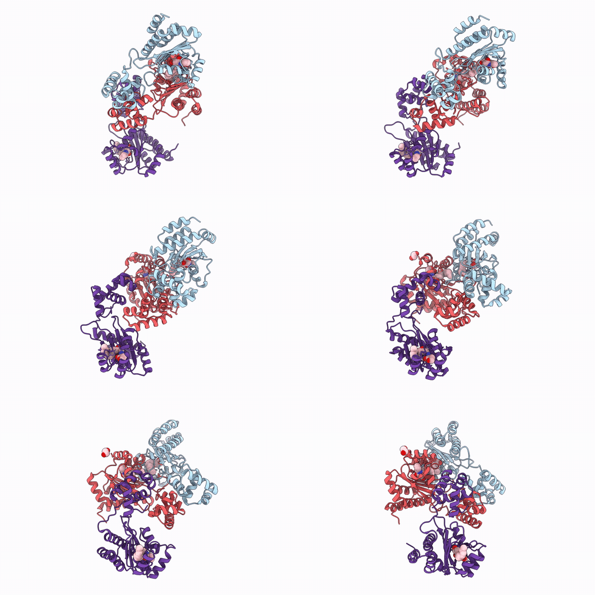
9ZZ0 image
Deposition Date 2026-01-06
Release Date 2026-09-23
Last Version Date 2026-10-14
Entry Detail
PDB ID:
9ZZ0
Keywords:
Title:
Crystal structure of MrtR bound to 3OH-C14 homoserine lactone
Biological Source:
Source Organism(s):
Expression System(s):
Method Details:
Experimental Method:
Resolution:
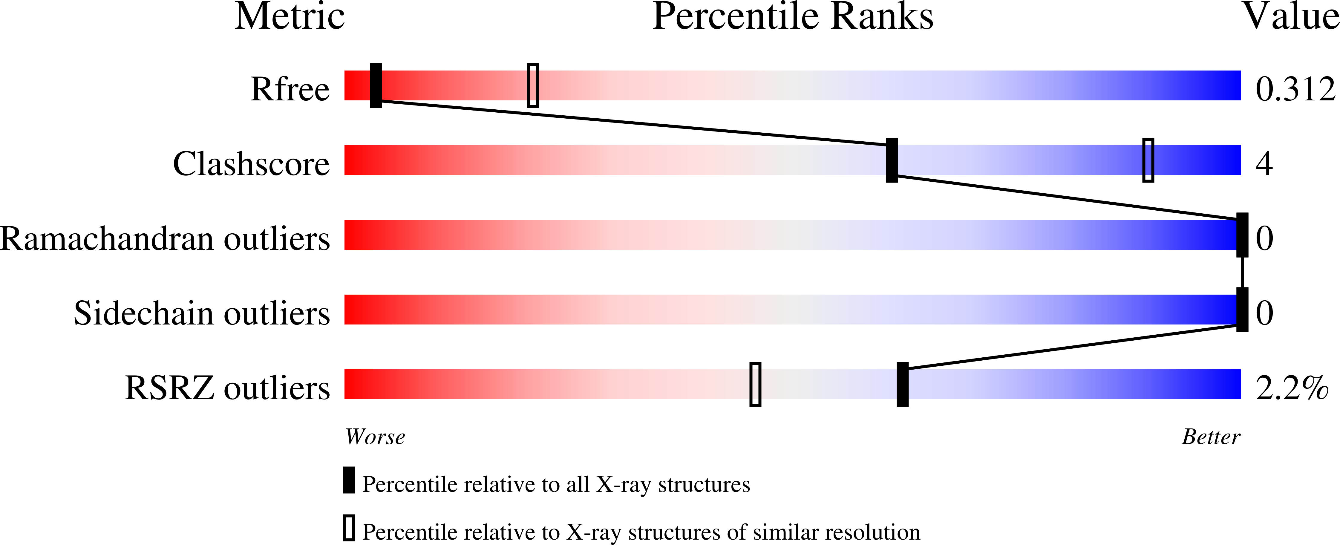
3.36 Å
R-Value Free:
0.30
R-Value Work:
0.25
R-Value Observed:
0.25
Space Group:
C 1 2 1
Macromolecular Entities
Polymer Type:polypeptide(L)
Molecule:MrtR
Gene (Uniprot):mrtR
Chain IDs:A, B, C
Chain Length:241
Number of Molecules:3
Biological Source:Mesorhizobium tianshanense
Primary Citation
MrtR of Mesorhizobium tianshanense reveals both activation and inhibition mechanisms of a LuxR-type quorum sensing receptor.
Proc.Natl.Acad.Sci.USA 123 e2616692123 e2616692123 (2026)
PMID: 42814558 DOI: 10.1073/pnas.2616692123

Abstact

Quorum sensing (QS) enables common gram-negative bacteria to coordinate collective behaviors through small molecule signals, yet how these signals tune receptor activity remains incompletely understood. Here, we define a mechanism by which ligand structure controls function in a LuxR-type QS receptor. Using structural and biochemical analyses, we investigate MrtR from Mesorhizobium tianshanense and show that ligand acyl-chain length governs receptor assembly and activity. We present full-length structures of MrtR bound to activating and inhibitory ligands, revealing a switch in oligomeric state. Long-chain (C14) N-acyl l-homoserine lactones (AHLs) act as agonists by promoting intra- and intersubunit interactions that lead to homodimerization and DNA binding. In contrast, shorter (C8) AHLs fail to promote these contacts, favoring a monomeric, inactive state. Ligands of intermediate length produce graded responses consistent with partial dimer stabilization. Biochemical measurements of DNA binding, thermostability, and oligomerization, together with targeted mutagenesis, support this model and establish the functional importance of key structural contacts. These findings provide a side-by-side structural comparison of a full-length LuxR-type receptor bound to both agonist and antagonist. Our results expand the known structural and mechanistic diversity of the LuxR family and suggest mechanistic similarities between structurally distinct receptors.

Legend

Protein

Chemical

Disease

Primary Citation of related structures
Feedback Form
Name
Email
Institute
Feedback