

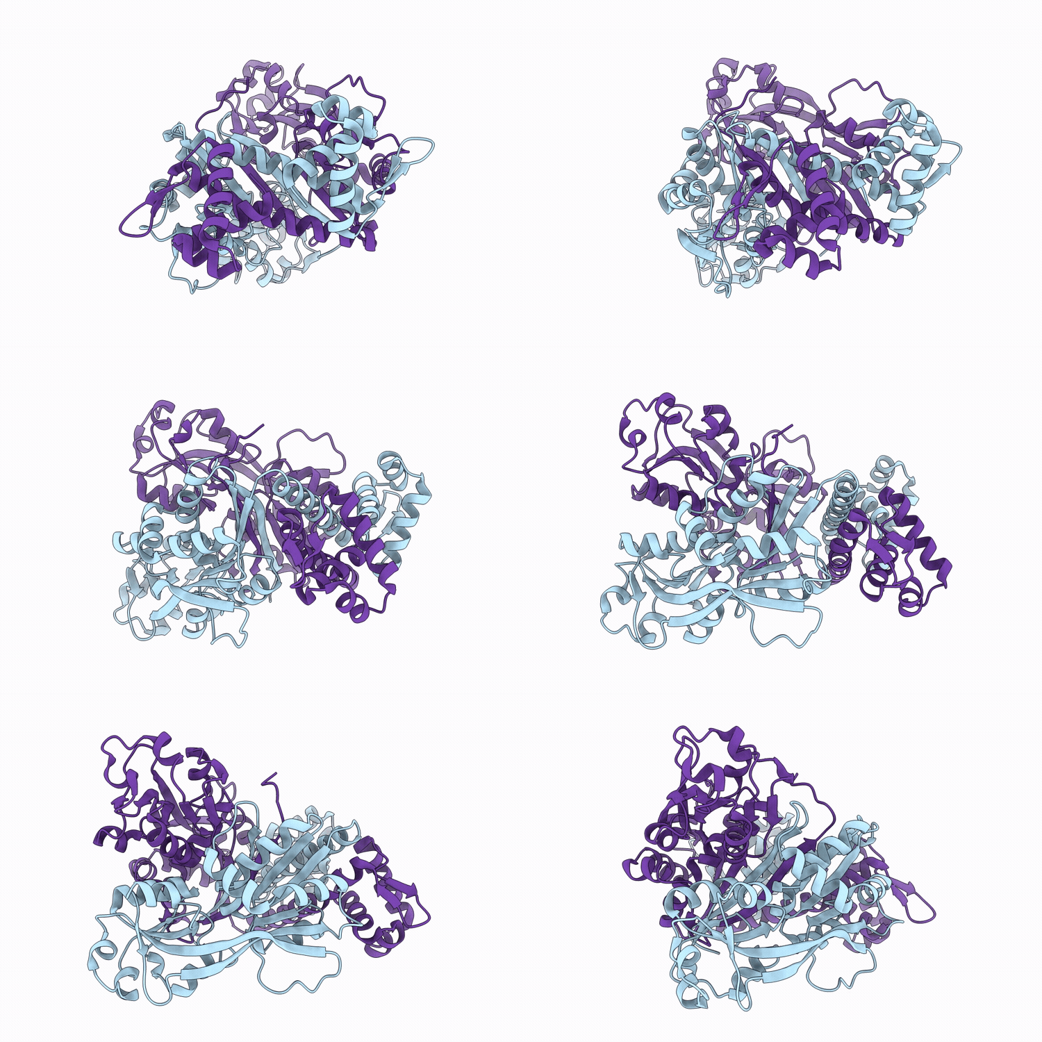
9VVT image
Deposition Date 2025-07-16
Release Date 2026-05-27
Last Version Date 2026-05-27
Entry Detail
PDB ID:
9VVT
Keywords:
Title:
Crystal structure of the LysR-type transcriptional regulator CutR from mycobacterium sp. strain JC1
Biological Source:
Source Organism(s):
Expression System(s):
Method Details:
Experimental Method:
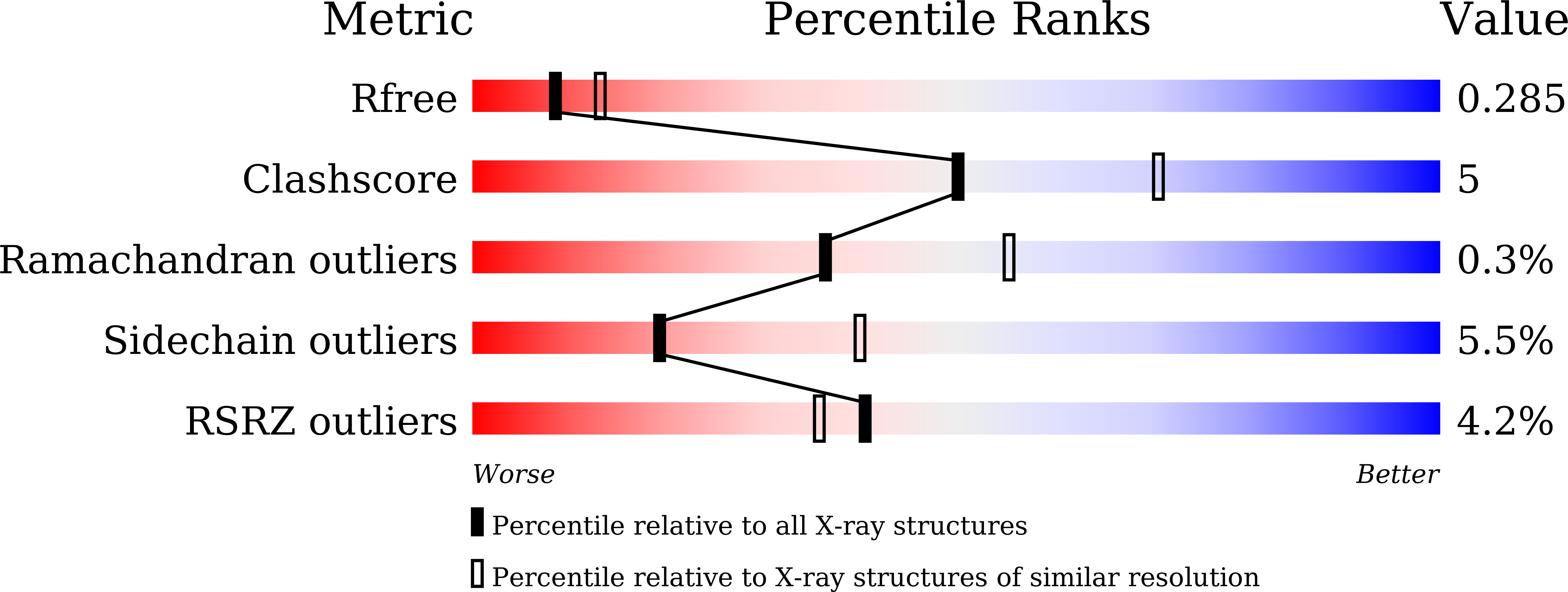
Resolution:
2.50 Å
R-Value Free:
0.26
R-Value Work:
0.21
R-Value Observed:
0.21
Space Group:
P 21 21 21
Macromolecular Entities
Structures with similar UniProt ID
Protein Blast
Polymer Type:polypeptide(L)
Molecule:LysR family transcriptional r
Gene (Uniprot):cutR
Chain IDs:A, B
Chain Length:326
Number of Molecules:2
Biological Source:Mycobacterium sp. DSM 3803
Modified Residue
Compound ID Chain ID Parent Comp ID Details 2D Image
MSE A MET modified residue
Primary Citation
Symmetric Dimeric Structure and Ligand Recognition of CutR, a LysR-Type Transcriptional Regulator from Mycobacterium sp. Strain JC1.
Int J Mol Sci 26 ? ? (2025)
PMID: 41226566 DOI: 10.3390/ijms262110533

Abstact

Mycobacteria possess carbon monoxide dehydrogenase (CO-DH) to utilize CO as an energy source and to resist host defense mechanisms. The expression of the CO-DH gene is regulated by CutR, a LysR-type transcriptional regulator (LTTR) that exhibits unique characteristics, suggesting that it functions as a dimer rather than the typical tetramer. Size-exclusion chromatography revealed that CutR forms a stable dimer. Electrophoretic mobility shift assays demonstrated that dimeric CutR specifically binds to an inverted repeat sequence (IR1) containing T-n12-A motifs located upstream of the cutB gene, which encodes the medium subunit of CO-DH. Crystal structure determination at 1.8 A resolution revealed that CutR consists of an N-terminal DNA-binding domain with a winged helix-turn-helix motif and a C-terminal ligand-binding domain comprising two regulatory subdomains (RD1 and RD2), forming a unique two-fold symmetrical homodimer. This dimer is stabilized through four interfaces, including an extensive 12-stranded antiparallel beta-sheet formed between RD1 subdomains via intertwining C-terminal beta11 strands. This represents the first symmetric dimeric LTTR structure with tightly associated ligand-binding domains. The recognition helices are spaced closer together than they are in typical DNA-bound LTTRs, despite binding longer T-n12-A sequences, suggesting that a conformational change is required to enhance DNA-binding affinity. A putative ligand-binding site was identified between the RD1 and RD2 subdomains, where glycerol binding induced local conformational changes. Comparative genomic analysis revealed conservation of CutR and the IR1 sequence across Mycobacterium species, supporting the dimeric regulatory mechanism and providing new insights into LTTR diversity.

Legend

Protein

Chemical

Disease

Primary Citation of related structures
Feedback Form
Name
Email
Institute
Feedback