

9SYU image
Deposition Date 2025-10-14
Release Date 2025-10-22
Last Version Date 2026-05-06
Entry Detail
PDB ID:
9SYU
Keywords:
Title:
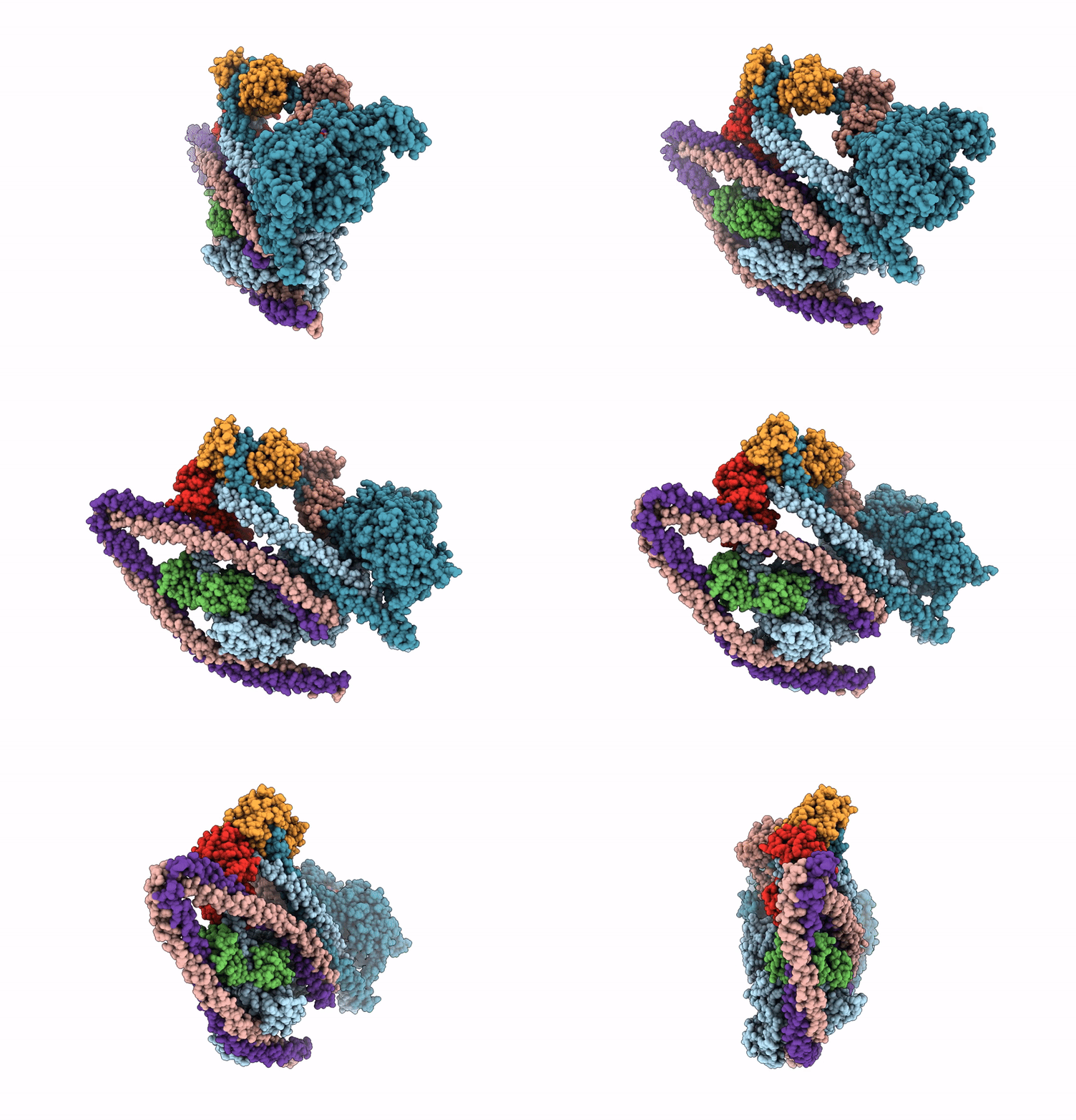
shutdown state non-muscle myosin 2A heads region
Biological Source:
Source Organism(s):
Homo sapiens (Taxon ID: 9606)
Gallus gallus (Taxon ID: 9031)
Bos taurus (Taxon ID: 9913)
Expression System(s):
Method Details:
Experimental Method:
Resolution:
2.98 Å
Aggregation State:
PARTICLE
Reconstruction Method:
SINGLE PARTICLE
Macromolecular Entities
Polymer Type:polypeptide(L)
Molecule:Isoform 1 of Myosin-9
Gene (Uniprot):MYH9
Chain IDs:A, B, G, H
Chain Length:1960
Number of Molecules:4
Biological Source:Homo sapiens
Polymer Type:polypeptide(L)
Molecule:Isoform 1 of Myosin light pol
Gene (Uniprot):MYL6
Chain IDs:C, D
Chain Length:151
Number of Molecules:2
Biological Source:Gallus gallus
Polymer Type:polypeptide(L)
Molecule:Myosin regulatory light polyp
Gene (Uniprot):MYL9
Chain IDs:E, F
Chain Length:172
Number of Molecules:2
Biological Source:Bos taurus
Primary Citation
Cryo-EM structure of shutdown human nonmuscle myosin 2A.
Sci Adv 12 eaed1858 eaed1858 (2026)
PMID: 41996489 DOI: 10.1126/sciadv.aed1858

Abstact

Determining the high-resolution structure of the widely expressed nonmuscle myosin 2A (NM2A), in its dephosphorylated shutdown state, is important in understanding its regulation and disease roles. In shutdown molecules, the coiled-coil tail wraps around the myosin heads, preventing them from forming filaments and binding to actin. We have solved the shutdown structure of NM2A to a global resolution of 3.0 angstroms in the head region and 6.3 angstroms for the whole molecule. This reveals specific ionic interactions that explain why the path of the coiled coil and the shutdown mechanism for NM2A differ from those of beta-cardiac myosin and provides key insight into how specific mutations likely destabilize the shutdown state, leading to disease.

Legend

Protein

Chemical

Disease

Primary Citation of related structures
Feedback Form
Name
Email
Institute
Feedback