Deposition Date
2025-08-12
Release Date
2026-03-18
Last Version Date
2026-03-18
Entry Detail
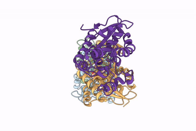
PDB ID:
9SD1
Keywords:
Title:
The R62Q clinical variant of human bisphosphoglycerate mutase (hBPGM).
Biological Source:
Source Organism(s):
Homo sapiens (Taxon ID: 9606)
Expression System(s):
Method Details:
Experimental Method:
Resolution:
2.50 Å
R-Value Free:
0.26
R-Value Work:
0.20
R-Value Observed:
0.21
Space Group:
P 1