

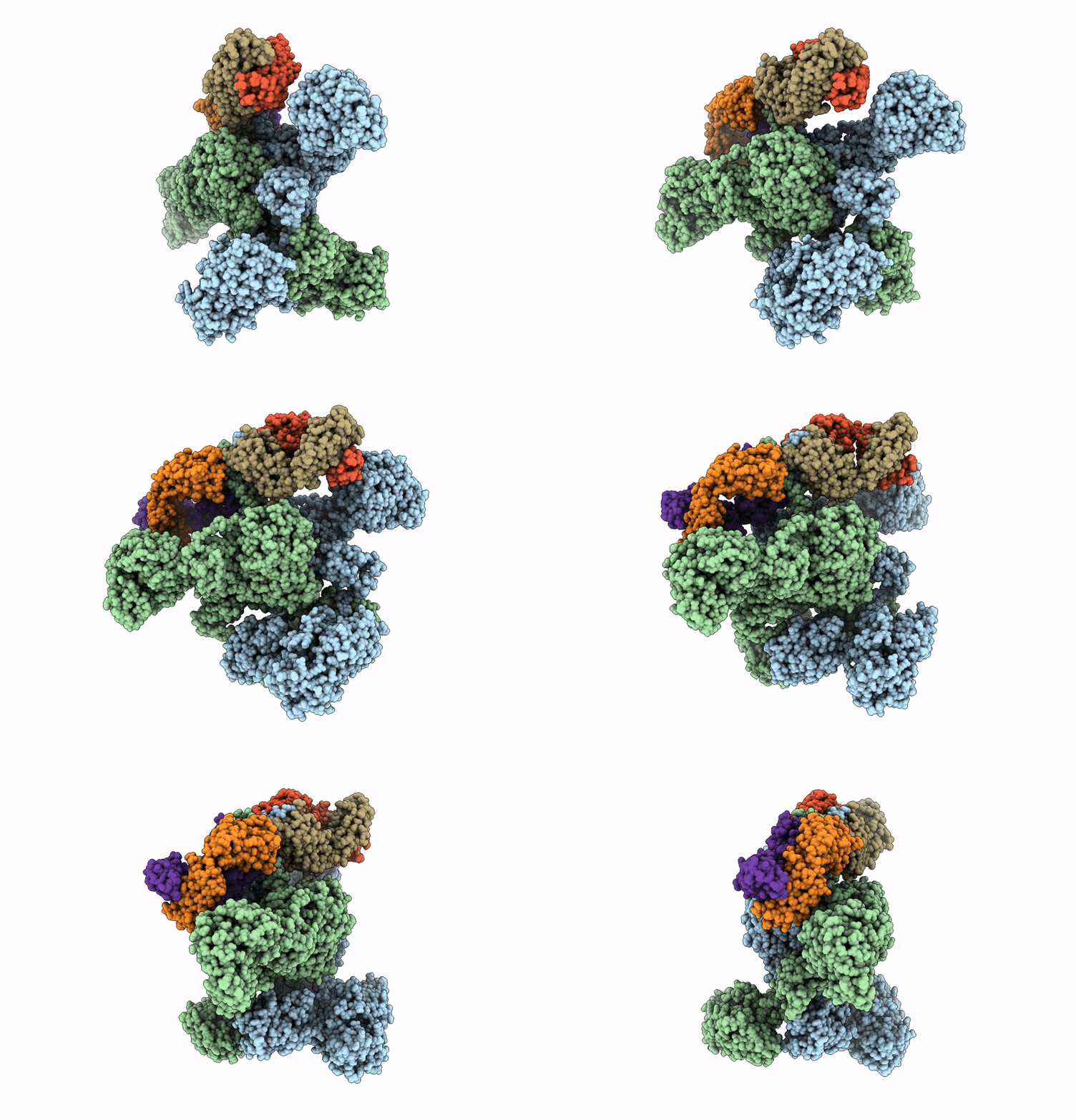
9PC6 image
Deposition Date 2025-06-27
Release Date 2025-07-23
Last Version Date 2026-09-30
Entry Detail
PDB ID:
9PC6
Title:
Antibody (1B2) Bound Crosslinked Rifamycin Synthetase Module 1 with a C-terminal Type II Thioesterase
Biological Source:
Source Organism(s):
Expression System(s):
Method Details:
Experimental Method:
Resolution:
3.96 Å
Aggregation State:
PARTICLE
Reconstruction Method:
SINGLE PARTICLE
Macromolecular Entities
Structural Superimposition Protein Blast
Polymer Type:polypeptide(L)
Molecule:6-deoxyerythronolide-B syntha
Gene (Uniprot):rifA, rifR
Chain IDs:A, B
Chain Length:1869
Number of Molecules:2
Biological Source:Amycolatopsis mediterranei
Polymer Type:polypeptide(L)
Molecule:Antibody Fragment 1B2 Light C
Chain IDs:E (auth: I), F (auth: L)
Chain Length:236
Number of Molecules:2
Biological Source:Homo sapiens
Polymer Type:polypeptide(L)
Molecule:Antibody Fragment 1B2 Heavy C
Chain IDs:C (auth: J), D (auth: K)
Chain Length:249
Number of Molecules:2
Biological Source:Homo sapiens
Modified Residue
Compound ID Chain ID Parent Comp ID Details 2D Image
4HH A, B SER modified residue
Ligand Molecules
Primary Citation
C-terminal dimerization motifs control asynchronous chain elongation during modular polyketide biosynthesis.
J.Biol.Chem. 302 113119 113119 (2026)
PMID: 42103224 DOI: 10.1016/j.jbc.2026.113119

Abstact

The rifamycin synthetase (RIFS) from the bacterium Amycolatopsis mediterranei is a homodimeric assembly line that catalyzes 40+ chemical reactions to generate a complex precursor of the antitubercular drug rifampicin. It consists of an N-terminal substrate loading module followed by a decamodular polyketide synthase (PKS). While the catalytic functions are known for each domain of RIFS, how these activities are spatially and temporally coordinated during polyketide assembly remains incompletely defined. Here, we address this problem with thiol-selective crosslinking to understand the basis for conformational asymmetry during polyketide chain elongation. Our data suggest that C-terminal dimerization motifs-which are ubiquitous in bacterial PKS assembly lines-force their adjacent substrate carrier protein (CP) domains to comigrate between two equivalent ketosynthase (KS) active site chambers. Cryogenic electron microscopy analysis of the first PKS module of RIFS further underscored this observation while revealing its unique architecture. Single-turnover kinetic analysis indicated that although changes to the C-terminus that reduced CP dimerization supported 2-fold greater KS:CP interactions, they were insufficient to overcome substoichiometric product accumulation on the homodimeric protein. Our findings illuminate factors underlying asymmetry during polyketide antibiotic biosynthesis and should be instructive to future megasynth(et)ase engineers.

Legend

Protein

Chemical

Disease

Primary Citation of related structures
Feedback Form
Name
Email
Institute
Feedback