9CX9 image
Deposition Date 2024-07-31
Release Date 2024-11-13
Last Version Date 2026-04-08
Entry Detail
PDB ID:
9CX9
Title:
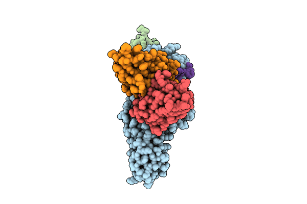
Structure of SH3 domain of Src in complex with beta-arrestin 1
Biological Source:
Source Organism(s):
Mus musculus (Taxon ID: 10090)
Rattus norvegicus (Taxon ID: 10116)
Gallus gallus (Taxon ID: 9031)
Homo sapiens (Taxon ID: 9606)
Method Details:
Experimental Method:
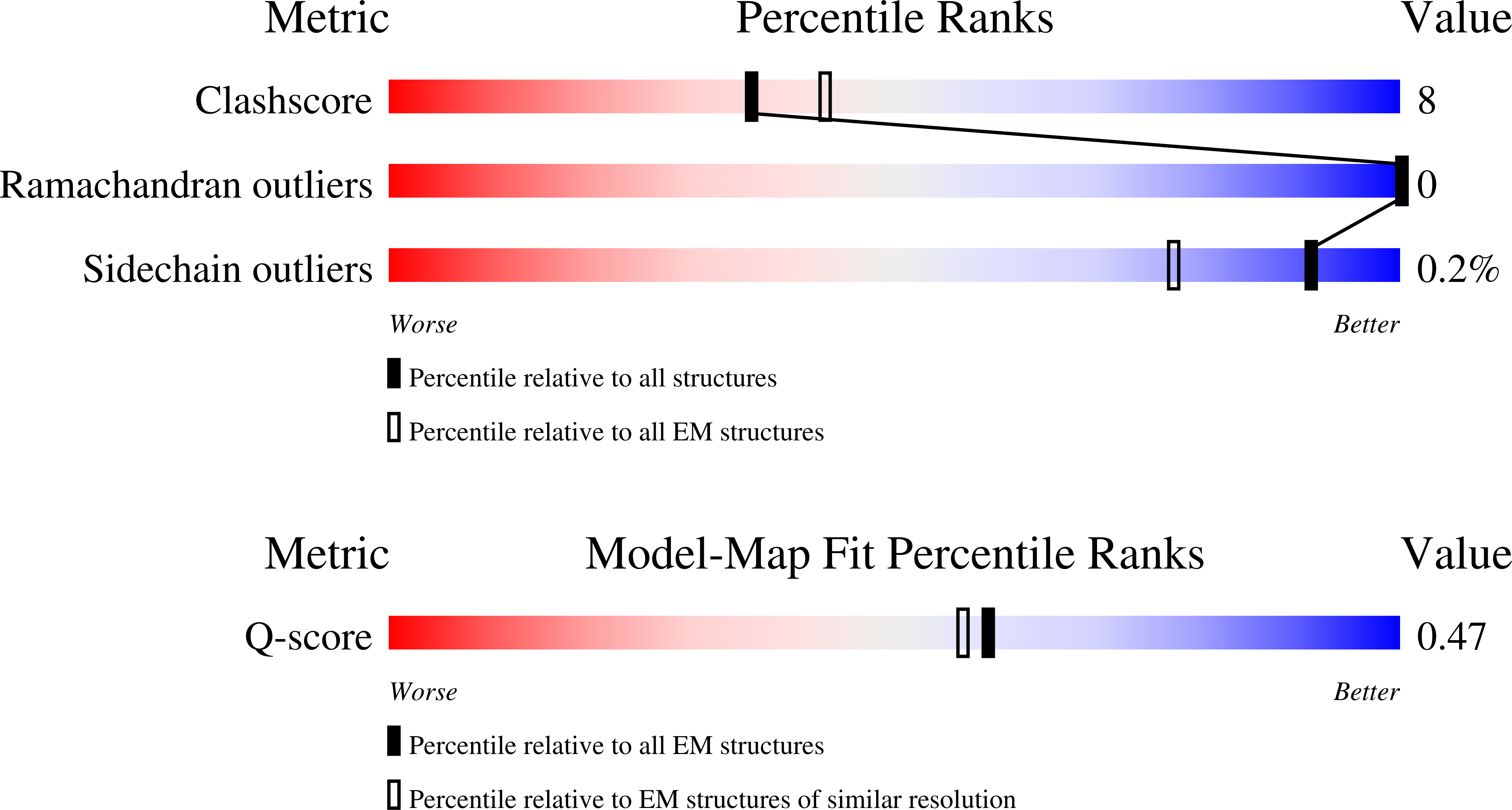
Resolution:
3.34 Å
Aggregation State:
PARTICLE
Reconstruction Method:
SINGLE PARTICLE
Macromolecular Entities
Structures with similar UniProt ID
Protein Blast
Polymer Type:polypeptide(L)
Molecule:Beta-arrestin-1
Gene (Uniprot):Arrb1
Mutagens:C59V, E92C, C125S, C140L, C150V, C242V, C251V, C269S
Chain IDs:D (auth: A)
Chain Length:392
Number of Molecules:1
Biological Source:Rattus norvegicus
Structures with similar UniProt ID
Protein Blast
Polymer Type:polypeptide(L)
Molecule:Proto-oncogene tyrosine-prote
Gene (Uniprot):SRC
Mutagens:R95C
Chain IDs:E (auth: B)
Chain Length:85
Number of Molecules:1
Biological Source:Gallus gallus
Protein Blast
Polymer Type:polypeptide(L)
Molecule:Antibody fragment Fab30, heav
Chain IDs:A (auth: H)
Chain Length:237
Number of Molecules:1
Biological Source:Mus musculus
Protein Blast
Polymer Type:polypeptide(L)
Molecule:Antibody fragment Fab30, ligh
Chain IDs:B (auth: L)
Chain Length:215
Number of Molecules:1
Biological Source:Mus musculus
Structures with similar UniProt ID
Protein Blast
Polymer Type:polypeptide(L)
Molecule:Vasopressin V2 receptor
Gene (Uniprot):AVPR2
Chain IDs:C (auth: V)
Chain Length:29
Number of Molecules:1
Biological Source:Homo sapiens
Modified Residue
Compound ID Chain ID Parent Comp ID Details 2D Image
SEP C SER modified residue
TPO C THR modified residue
Ligand Molecules
Primary Citation
Mechanism of beta-arrestin 1 mediated Src activation via Src SH3 domain revealed by cryo-electron microscopy.
Nat Commun 17 ? ? (2026)
PMID: 41720803 DOI: 10.1038/s41467-026-69884-1

Abstact

Beta-arrestins (betaarrs) are key regulators and transducers of G-protein coupled receptor signaling; however, little is known of how betaarrs communicate with their downstream effectors. Here, we delineate structural mechanisms underlying betaarr-mediated signal transduction. Using cryo-electron microscopy, we elucidate how betaarr1 recruits and activates the non-receptor tyrosine kinase Src, a well-established signaling partner of betaarrs. betaarr1 engages Src SH3 through two distinct sites, each employing a different recognition mechanism: a polyproline motif in the N-domain and a non-proline-based interaction in the central crest region. At both sites betaarr1 interacts with the aromatic surface of SH3, disrupting the autoinhibited conformation of Src and directly triggering its allosteric activation. This structural evidence establishes betaarr1 as an active regulatory protein rather than a passive scaffold and suggests a potentially general mechanism for betaarr-mediated signaling across diverse effectors.

Legend

Protein

Chemical

Disease

Primary Citation of related structures
Feedback Form
Name
Email
Institute
Feedback