

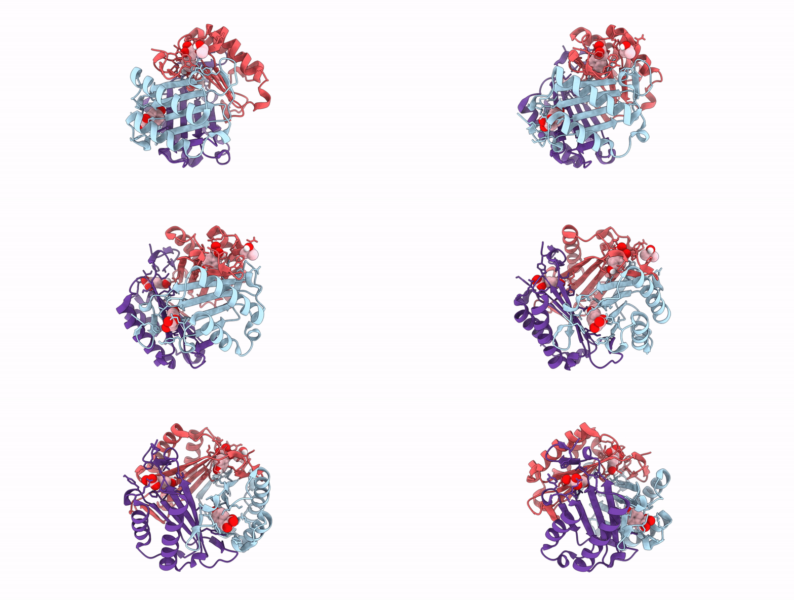
8TT8 image
Deposition Date 2023-08-13
Release Date 2025-08-27
Last Version Date 2026-09-09
Entry Detail
PDB ID:
8TT8
Keywords:
Title:
Joint Xray/Neutron structure of Macrophage Migration Inhibitory Factor (MIF) Bound to 4-hydroxyphenylpyruvate at room temperature
Biological Source:
Source Organism(s):
Homo sapiens (Taxon ID: 9606)
Expression System(s):
Method Details:
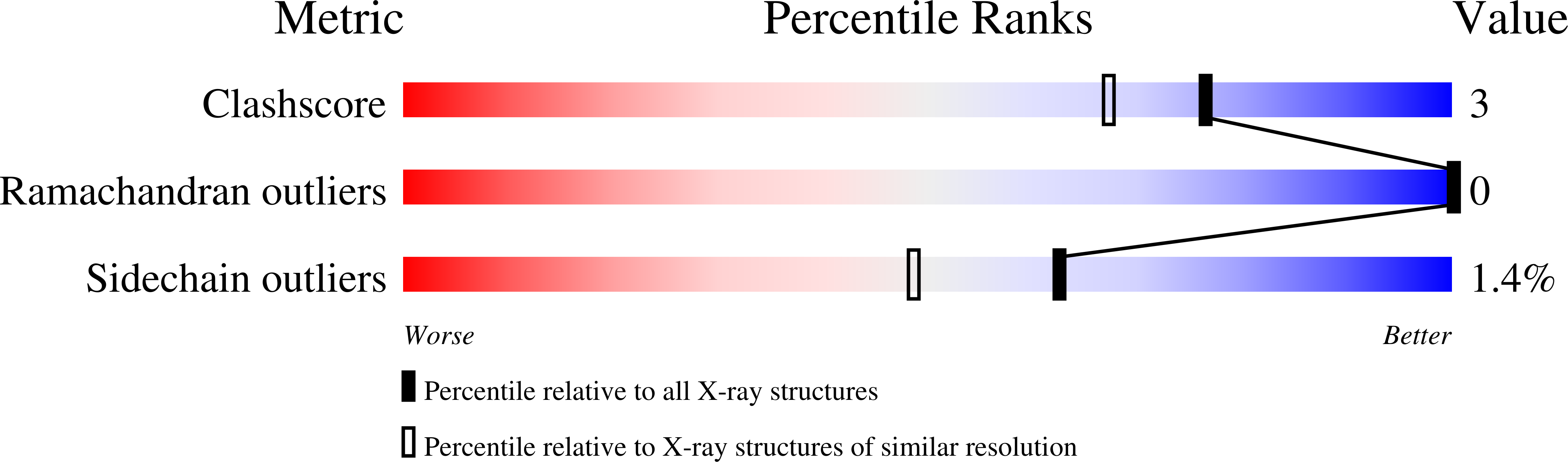
R-Value Free:
['0.18
R-Value Work:
['0.15
R-Value Observed:
['0.15
Space Group:
P 21 21 21
Macromolecular Entities
Polymer Type:polypeptide(L)
Molecule:Macrophage migration inhibito
Gene (Uniprot):MIF
Chain IDs:A, B, C
Chain Length:114
Number of Molecules:3
Biological Source:Homo sapiens
Primary Citation
Identification of a key water molecule involved in the macrophage migration inhibitory factor-catalyzed tautomerization of para-hydroxyphenylpyruvate using neutron crystallography.
Protein Sci. 35 e70666 e70666 (2026)
PMID: 42568349 DOI: 10.1002/pro.70666

Abstact

Neutron crystallography was used to determine a 2.5-A resolution all-atom structure of macrophage migration inhibitory factor (MIF) interacting with 3-(4-hydroxyphenyl)-pyruvate (HPP). MIF is a pro-inflammatory, pro-tumorigenic protein that may be an attractive therapeutic target. MIF catalyzes the interconversion of the keto and enol forms of HPP by a tautomerase reaction. Although HPP is evidently not a physiological substrate of MIF, many compounds that inhibit this activity in enzymatic assays have been found also to inhibit physiological activities of MIF. Therefore, the MIF-catalyzed HPP tautomerization reaction is used in initial screening of compounds in the search for inhibitors of MIF physiological activity. The neutron diffraction-derived crystal structure reveals the position of a water molecule involved in the tautomerization reaction, and also confirms the charged state of lysine-32 in the active site. The structure confirms the previously proposed catalytic mechanism of MIF, with the N-terminal Pro-1 abstracting a proton to generate an HPP enolate intermediate which is subsequently protonated. The structure reported herein reveals that this proton is supplied by a neighboring water molecule. Along with the neutron structure, a room-temperature synchrotron x-ray crystal structure reveals a covalent adduct between HPP and MIF. While this adduct is a result of radiation-induced chemistry, its formation confirms the catalytic role of the active site residue because a covalent complex could only form if the reactive carbon of the substrate is correctly positioned by the enzyme.

Legend

Protein

Chemical

Disease

Primary Citation of related structures
Feedback Form
Name
Email
Institute
Feedback