

5XGH image
Deposition Date 2017-04-13
Release Date 2018-04-25
Last Version Date 2024-03-27
Entry Detail
PDB ID:
5XGH
Title:
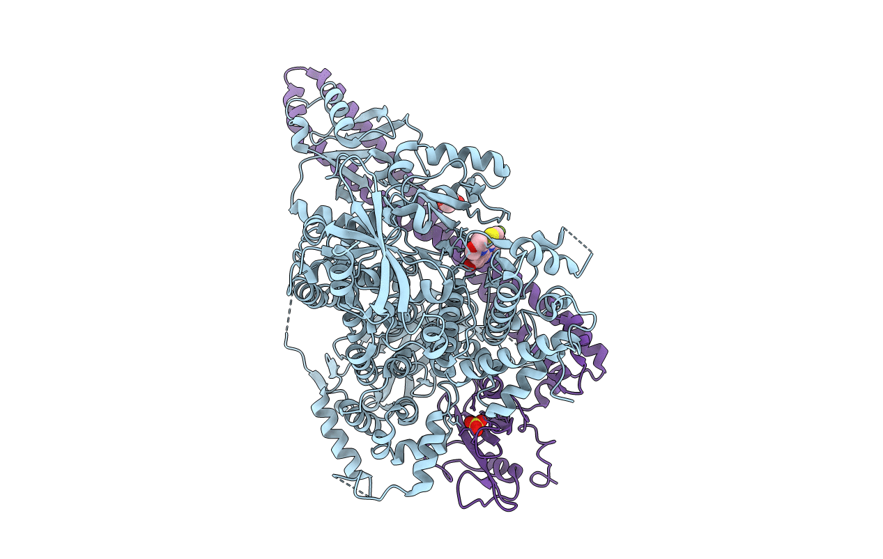
Crystal structure of PI3K complex with an inhibitor
Biological Source:
Source Organism(s):
Homo sapiens (Taxon ID: 9606)
Expression System(s):
Method Details:
Experimental Method:
Resolution:
2.97 Å
R-Value Free:
0.28
R-Value Work:
0.23
R-Value Observed:
0.23
Space Group:
P 21 21 21
Macromolecular Entities
Polymer Type:polypeptide(L)
Molecule:Phosphatidylinositol 4,5-bisp
Gene (Uniprot):PIK3CA
Chain IDs:A
Chain Length:1048
Number of Molecules:1
Biological Source:Homo sapiens
Polymer Type:polypeptide(L)
Molecule:Phosphatidylinositol 3-kinase
Gene (Uniprot):PIK3R1
Chain IDs:B
Chain Length:277
Number of Molecules:1
Biological Source:Homo sapiens
Primary Citation
New Insights into PI3K Inhibitor Design using X-ray Structures of PI3K alpha Complexed with a Potent Lead Compound.
Sci Rep 7 14572 14572 (2017)
PMID: 29109464 DOI: 10.1038/s41598-017-15260-5

Abstact

Phosphatidylinositol 3-kinase α is an attractive target to potentially treat a range of cancers. Herein, we described the evolution of a reported PI3K inhibitor into a moderate PI3Kα inhibitor with a low molecular weight. We used X-ray crystallography to describe the accurate binding mode of the compound YXY-4F. A comparison of the p110α-YXY-4F and apo p110α complexes showed that YXY-4F induced additional space by promoting a flexible conformational change in residues Ser773 and Ser774 in the PI3Kα ATP catalytic site. Specifically, residue 773(S) in PI3Kα is quite different from that of PI3Kβ (D), γ (A), and δ (D), which might guide further optimization of substituents around the NH group and phenyl group to improve the selectivity and potency of PI3Kα.

Legend

Protein

Chemical

Disease

Primary Citation of related structures
Feedback Form
Name
Email
Institute
Feedback