

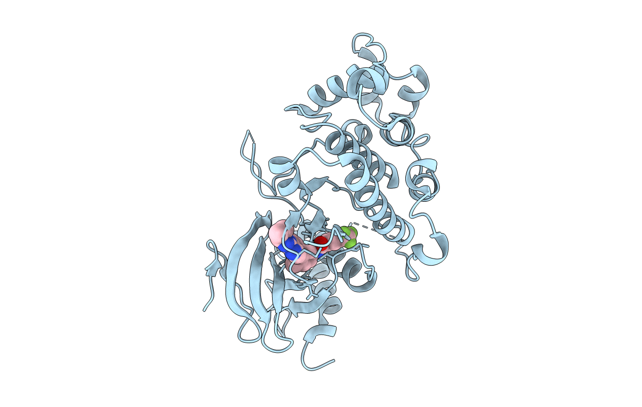
4I5H image
Deposition Date 2012-11-28
Release Date 2013-07-10
Last Version Date 2024-02-28
Entry Detail
PDB ID:
4I5H
Title:
Crystal Structure of a Double Mutant Rat Erk2 Complexed With a Type II Quinazoline Inhibitor
Biological Source:
Source Organism(s):
Expression System(s):
Method Details:
Experimental Method:
Resolution:
1.90 Å
R-Value Free:
0.25
R-Value Work:
0.21
R-Value Observed:
0.22
Space Group:
P 31 2 1

Macromolecular Entities
Polymer Type:polypeptide(L)
Molecule:Mitogen-activated protein kin
Gene (Uniprot):Mapk1
Mutagens:Q103A, C164L
Chain IDs:A
Chain Length:359
Number of Molecules:1
Biological Source:Rattus norvegicus
Ligand Molecules
Primary Citation
Sequence determinants of a specific inactive protein kinase conformation.
Chem. Biol. 20 806 815 (2013)
PMID: 23790491 DOI: 10.1016/j.chembiol.2013.05.005

Abstact

Only a small percentage of protein kinases have been shown to adopt a distinct inactive ATP-binding site conformation, called the Asp-Phe-Gly-out (DFG-out) conformation. Given the high degree of homology within this enzyme family, we sought to understand the basis of this disparity on a sequence level. We identified two residue positions that sensitize mitogen-activated protein kinases (MAPKs) to inhibitors that stabilize the DFG-out inactive conformation. After characterizing the structure and dynamics of an inhibitor-sensitive MAPK mutant, we demonstrated the generality of this strategy by sensitizing a kinase (apoptosis signal-regulating kinase 1) not in the MAPK family to several DFG-out stabilizing ligands, using the same residue positions. The use of specific inactive conformations may aid the study of noncatalytic roles of protein kinases, such as binding partner interactions and scaffolding effects.

Legend

Protein

Chemical

Disease

Primary Citation of related structures
Feedback Form
Name
Email
Institute
Feedback