

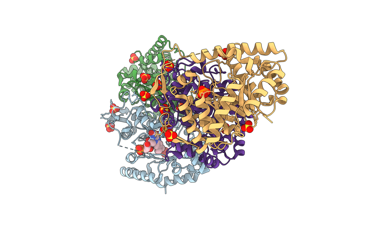
3FGC image
Deposition Date 2008-12-05
Release Date 2009-05-26
Last Version Date 2023-09-06
Entry Detail
PDB ID:
3FGC
Keywords:
Title:
Crystal Structure of the Bacterial Luciferase:Flavin Complex Reveals the Basis of Intersubunit Communication
Biological Source:
Source Organism(s):
Vibrio harveyi (Taxon ID: 669)
Expression System(s):
Method Details:
Experimental Method:
Resolution:
2.30 Å
R-Value Free:
0.24
R-Value Work:
0.18
R-Value Observed:
0.18
Space Group:
P 21 21 21
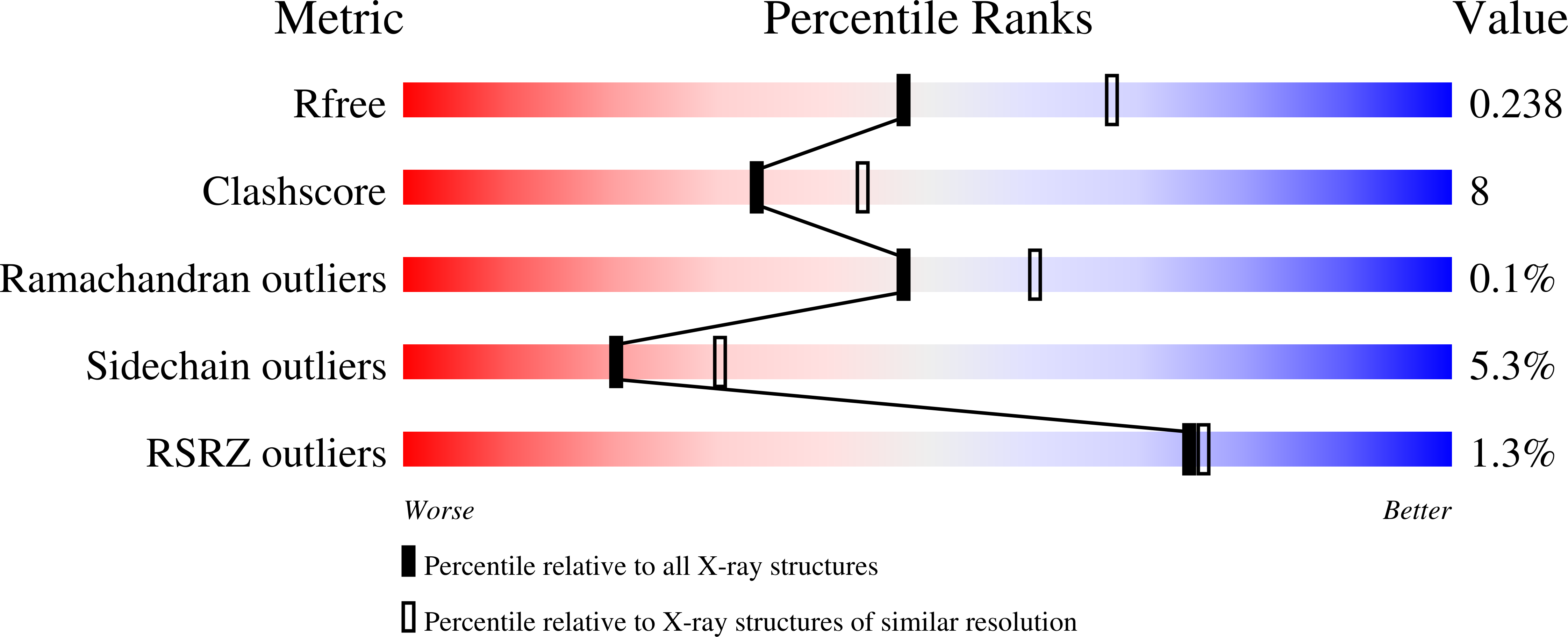
Macromolecular Entities
Polymer Type:polypeptide(L)
Molecule:Alkanal monooxygenase alpha c
Gene (Uniprot):luxA
Chain IDs:A, C
Chain Length:355
Number of Molecules:2
Biological Source:Vibrio harveyi
Polymer Type:polypeptide(L)
Molecule:Alkanal monooxygenase beta ch
Gene (Uniprot):luxB
Chain IDs:B, D
Chain Length:332
Number of Molecules:2
Biological Source:Vibrio harveyi
Primary Citation
Crystal structure of the bacterial luciferase/flavin complex provides insight into the function of the beta subunit.
Biochemistry 48 6085 6094 (2009)
PMID: 19435287 DOI: 10.1021/bi900003t

Abstact

Bacterial luciferase from Vibrio harveyi is a heterodimer composed of a catalytic alpha subunit and a homologous but noncatalytic beta subunit. Despite decades of enzymological investigation, structural evidence defining the active center has been elusive. We report here the crystal structure of V. harveyi luciferase bound to flavin mononucleotide (FMN) at 2.3 A. The isoalloxazine ring is coordinated by an unusual cis-Ala-Ala peptide bond. The reactive sulfhydryl group of Cys106 projects toward position C-4a, the site of flavin oxygenation. This structure also provides the first data specifying the conformations of a mobile loop that is crystallographically disordered in both prior crystal structures [(1995) Biochemistry 34, 6581-6586; (1996) J. Biol. Chem. 271, 21956 21968]. This loop appears to be a boundary between solvent and the active center. Within this portion of the protein, a single contact was observed between Phe272 of the alpha subunit, not seen in the previous structures, and Tyr151 of the beta subunit. Substitutions at position 151 on the beta subunit caused reductions in activity and total quantum yield. Several of these mutants were found to have decreased affinity for reduced flavin mononucleotide (FMNH(2)). These findings partially address the long-standing question of how the beta subunit stabilizes the active conformation of the alpha subunit, thereby participating in the catalytic mechanism.

Legend

Protein

Chemical

Disease

Primary Citation of related structures
Feedback Form
Name
Email
Institute
Feedback