

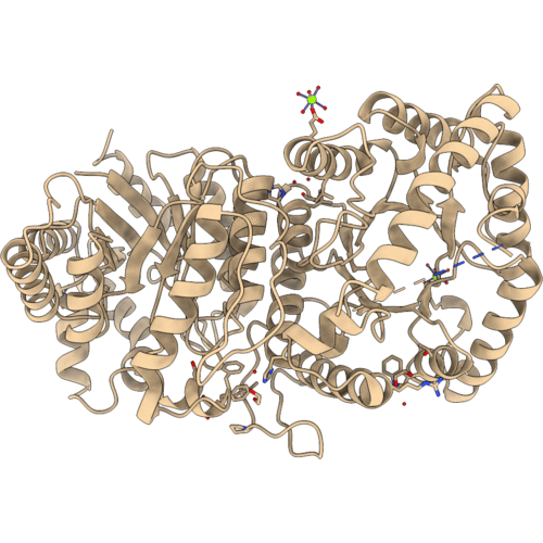
1LUC image
Deposition Date 1996-05-10
Release Date 1996-12-07
Last Version Date 2024-02-14
Entry Detail
PDB ID:
1LUC
Keywords:
Title:
BACTERIAL LUCIFERASE
Biological Source:
Source Organism(s):
Vibrio harveyi (Taxon ID: 669)
Expression System(s):
Method Details:

Experimental Method:
Resolution:
1.50 Å
Space Group:
C 1 2 1
Macromolecular Entities
Polymer Type:polypeptide(L)
Molecule:BACTERIAL LUCIFERASE
Gene (Uniprot):luxA
Chain IDs:A
Chain Length:355
Number of Molecules:1
Biological Source:Vibrio harveyi
Polymer Type:polypeptide(L)
Molecule:BACTERIAL LUCIFERASE
Gene (Uniprot):luxB
Chain IDs:B
Chain Length:324
Number of Molecules:1
Biological Source:Vibrio harveyi
Primary Citation
The 1.5-A resolution crystal structure of bacterial luciferase in low salt conditions.
J. Biol. Chem. 271 21956 21968 (1996)
PMID: 8703001 DOI: 10.1074/jbc.271.36.21956

Abstact

Bacterial luciferase is a flavin monooxygenase that catalyzes the oxidation of a long-chain aldehyde and releases energy in the form of visible light. A new crystal form of luciferase cloned from Vibrio harveyi has been grown under low-salt concentrations, which diffract x-rays beyond 1.5-A resolution. The x-ray structure of bacterial luciferase has been refined to a conventional R-factor of 18.2% for all recorded synchrotron data between 30.0 and 1.50-A resolution. Bacterial luciferase is an alpha-beta heterodimer, and the individual subunits fold into a single domain (beta/alpha)8 barrel. The high resolution structure reveals a non-prolyl cis peptide bond that forms between Ala74 and Ala75 in the alpha subunit near the putative active site. This cis peptide bond may have functional significance for creating a cavity at the active site. Bacterial luciferase employs reduced flavin as a substrate rather than a cofactor. The structure presented was determined in the absence of substrates. A comparison of the structural similarities between luciferase and a nonfluorescent flavoprotein, which is expressed in the lux operon of one genus of bioluminescent bacteria, suggests that the two proteins originated from a common ancestor. However, the flavin binding sites of the nonfluorescent protein are likely not representative of the flavin binding site on luciferase. The structure presented here will furnish a detailed molecular model for all bacterial luciferases.

Legend

Protein

Chemical

Disease

Primary Citation of related structures
Feedback Form
Name
Email
Institute
Feedback