

37UU image
Deposition Date 2026-08-06
Release Date 2026-11-04
Last Version Date 2026-11-04
Entry Detail
PDB ID:
37UU
Keywords:
Title:
Cryo-EM structure of human Fibroblast Activation Protein alpha in complex with F7 VHH
Biological Source:
Source Organism(s):
Camelidae (Taxon ID: 9835)
Homo sapiens (Taxon ID: 9606)
Expression System(s):
Method Details:
Experimental Method:
Resolution:
2.50 Å
Aggregation State:
PARTICLE
Reconstruction Method:
SINGLE PARTICLE
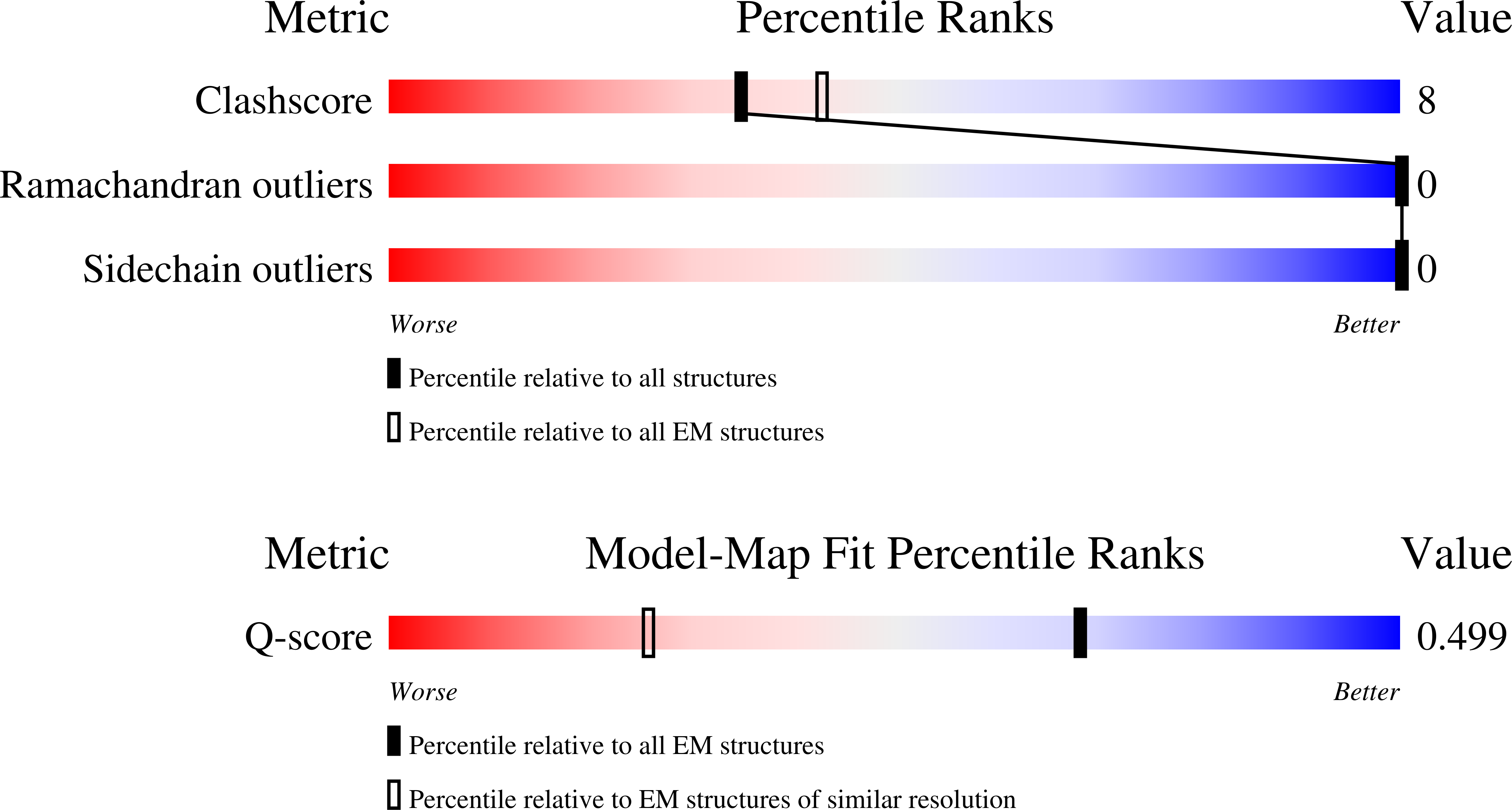
Macromolecular Entities
Polymer Type:polypeptide(L)
Molecule:Fibroblast activation protein
Gene (Uniprot):FAP
Chain IDs:B (auth: A), D (auth: B)
Chain Length:753
Number of Molecules:2
Biological Source:Homo sapiens
Polymer Type:polypeptide(L)
Molecule:F7 VHH-human IgG1 Fc fusion p
Chain IDs:A (auth: C), C (auth: D)
Chain Length:358
Number of Molecules:2
Biological Source:Camelidae
Ligand Molecules
Primary Citation
A shark variable new antigen receptor recognizes an occluded epitope of fibroblast activation protein.
Biorxiv ? ? ? (2026)
PMID: 42845595 DOI: 10.64898/2026.08.11.744296

Abstact

Variable new antigen receptors (VNARs) are the smallest naturally occurring antibody binding domains. Their size allows VNARs to access sterically restricted epitopes that are inaccessible to conventional antibodies. We recently identified a suite of VNARs that target fibroblast activation protein (FAP), a stromal serine protease indicative of extracellular matrix remodeling. The presence of FAP on the surface of cancer-associated fibroblasts (CAFs) that promote immunosuppression has made FAP a compelling therapeutic target for cancer therapy. Although antibodies targeting FAP have been developed, there is a paucity of information on how biologics engage FAP. Here, we used single-particle cryogenic electron microscopy (cryo-EM) to compare FAP recognition of three antibody architectures: a shark-derived VNAR, variable heavy (V(H)) and light domains (V(L)) of a humanized Immunoglobulin G (IgG), and a camelid-derived VHH. The humanized V(H)-V(L) domains and camelid VHH both target a solvent-exposed beta-propeller domain, whereas the VNAR binds a highly conserved, topologically recessed epitope at the FAP dimer interface. Radical-footprinting mass spectrometry (MS) further mapped two additional immune-derived VNARs to distinct FAP surfaces outside the shared beta-propeller epitope. These findings demonstrate how unique VNAR architecture can expand access to underexplored FAP surfaces and establish a structural framework for rational multiepitope targeting strategies.

Legend

Protein

Chemical

Disease

Primary Citation of related structures
Feedback Form
Name
Email
Institute
Feedback