

37JU image
Deposition Date 2026-07-23
Release Date 2026-10-28
Last Version Date 2026-10-28
Entry Detail
PDB ID:
37JU
Keywords:
Title:
PTDSS2 structure in EDTA
Biological Source:
Source Organism(s):
Equus caballus (Taxon ID: 9796)
Expression System(s):
Method Details:
Experimental Method:
Resolution:
2.80 Å
Aggregation State:
PARTICLE
Reconstruction Method:
SINGLE PARTICLE
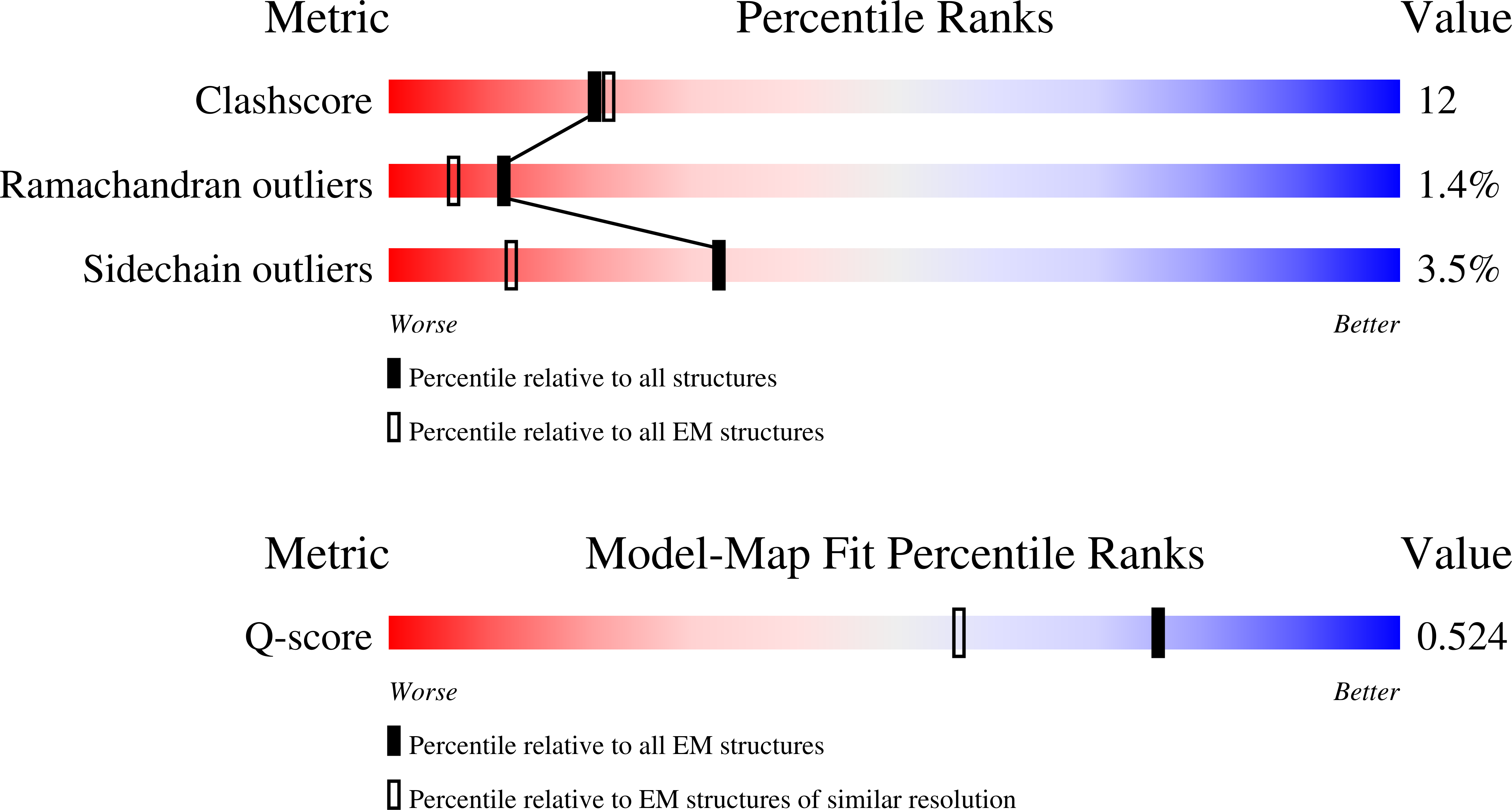
Macromolecular Entities
Polymer Type:polypeptide(L)
Molecule:Phosphatidylserine synthase
Gene (Uniprot):PTDSS2
Chain IDs:A, B (auth: C)
Chain Length:498
Number of Molecules:2
Biological Source:Equus caballus
Primary Citation
Mechanisms of substrate recognition and catalysis in a mammalian phosphatidylserine synthase 2.
Proc.Natl.Acad.Sci.USA 123 e2607382123 e2607382123 (2026)
PMID: 42809401 DOI: 10.1073/pnas.2607382123

Abstact

Mammalian phosphatidylserine synthase-1 and -2, PSS1 and PSS2, synthesize phosphatidylserine (PS) by replacing the headgroup of either phosphatidylcholine or phosphatidylethanolamine (PE) with a serine. We determined structures of PSS2 from Equus caballus in complex with either a PE or a serine substrate to resolutions of 2.8 to 3.2 A. The structures define substrate binding sites and reveal that the phosphate group of the PE is coordinated by two Ca(2+). In addition, we found that PSS2 has significant phospholipase D (PLD) activity in the absence of serine and that Ca(2+) is required for the PLD activity. These findings enrich and refine our knowledge in the mechanisms of substrate recognition and catalysis in mammalian PSS.

Legend

Protein

Chemical

Disease

Primary Citation of related structures
Feedback Form
Name
Email
Institute
Feedback