

35SG image
Deposition Date 2026-05-14
Release Date 2026-09-16
Last Version Date 2026-09-30
Entry Detail
PDB ID:
35SG
Title:
VIPR Binary Complex with two filaments
Biological Source:
Source Organism(s):
Expression System(s):
Method Details:
Experimental Method:
Resolution:
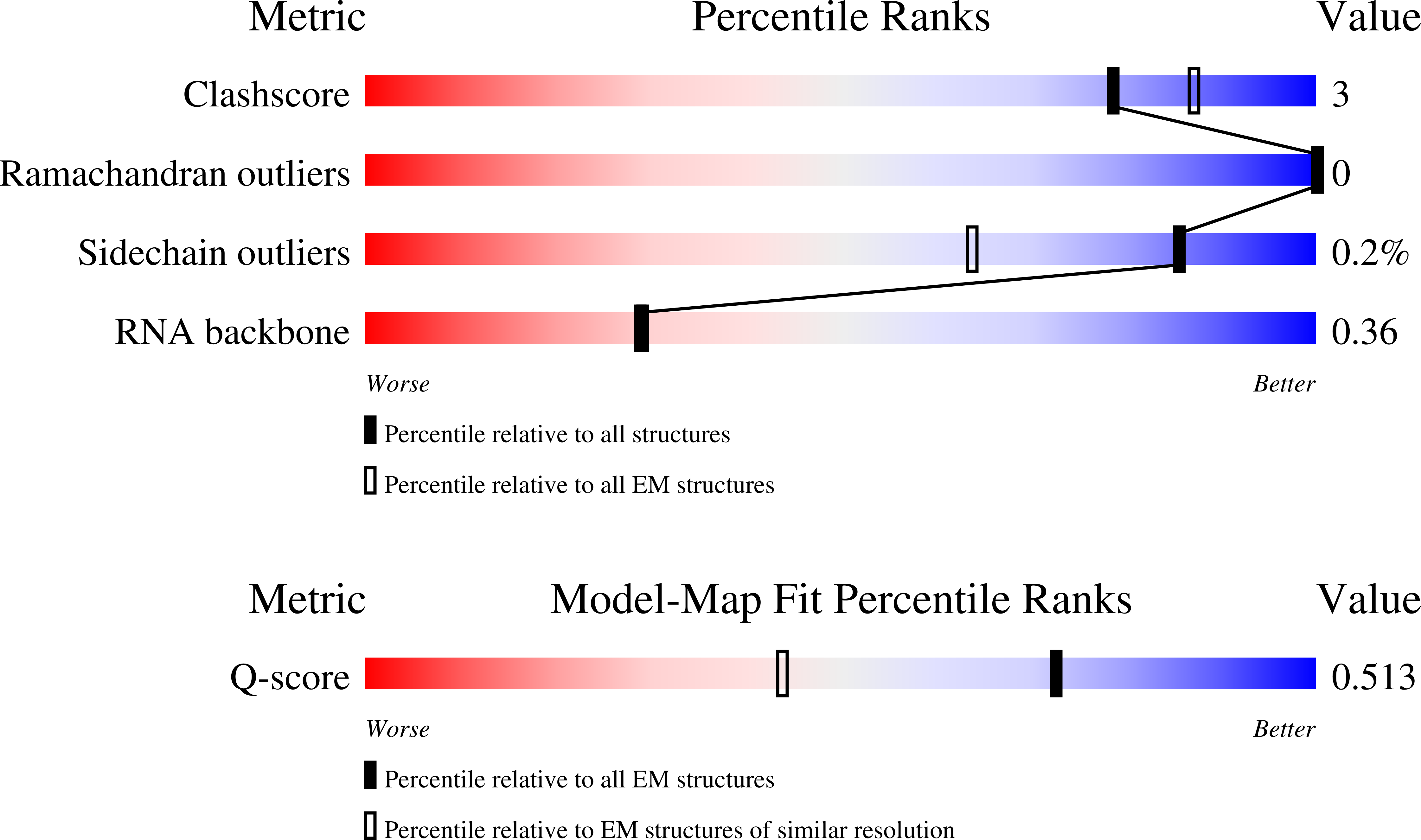
2.70 Å
Aggregation State:
PARTICLE
Reconstruction Method:
SINGLE PARTICLE
Macromolecular Entities
Polymer Type:polyribonucleotide
Molecule:vrRNA
Chain IDs:H (auth: A)
Chain Length:94
Number of Molecules:1
Biological Source:Suspvirus SUSP1
Structural Superimposition Protein Blast
Polymer Type:polypeptide(L)
Molecule:Vipr Protein
Chain IDs:A (auth: J), B (auth: I), C (auth: H), D (auth: G), E (auth: F), F (auth: E), G (auth: D)
Chain Length:202
Number of Molecules:7
Biological Source:Suspvirus SUSP1
Ligand Molecules
Primary Citation
VIPR RNA-guided DNA recognition by noncontiguous geometric triplex formation.
Science 393 1236 1240 (2026)
PMID: 42752121 DOI: 10.1126/science.aei3472

Abstact

Viral interference programmable repeat (VIPR) systems use a noncontiguous code for RNA-guided transcriptional silencing. How the Vipr protein and a VIPR RNA (vrRNA) comprising alternating GGY and NN segments achieve precise DNA targeting is unknown. Here, we present 21 cryo-electron microscopy structures that help explain the mechanism of target engagement. Vipr protomers oligomerize along the vrRNA to form a right-handed helical filament, sequestering each GGY motif and positioning the adjacent NN bases for target base pairing. DNA binding, in which every third nucleotide is skipped, results in a gapped vrRNA-DNA hybrid helix that encircles the nontarget DNA strand to form a geometric triplex. These findings suggest that triplex-mediated target-strand handoff could enable noncontiguous and programmable RNA-guided DNA recognition in VIPR systems.

Legend

Protein

Chemical

Disease

Primary Citation of related structures
Feedback Form
Name
Email
Institute
Feedback