

30HL image
Deposition Date 2026-04-26
Release Date 2026-11-04
Last Version Date 2026-11-11
Entry Detail
PDB ID:
30HL
Keywords:
Title:
Structure of inhibitor bound PriA
Biological Source:
Source Organism(s):
Expression System(s):
Method Details:
Experimental Method:
Resolution:
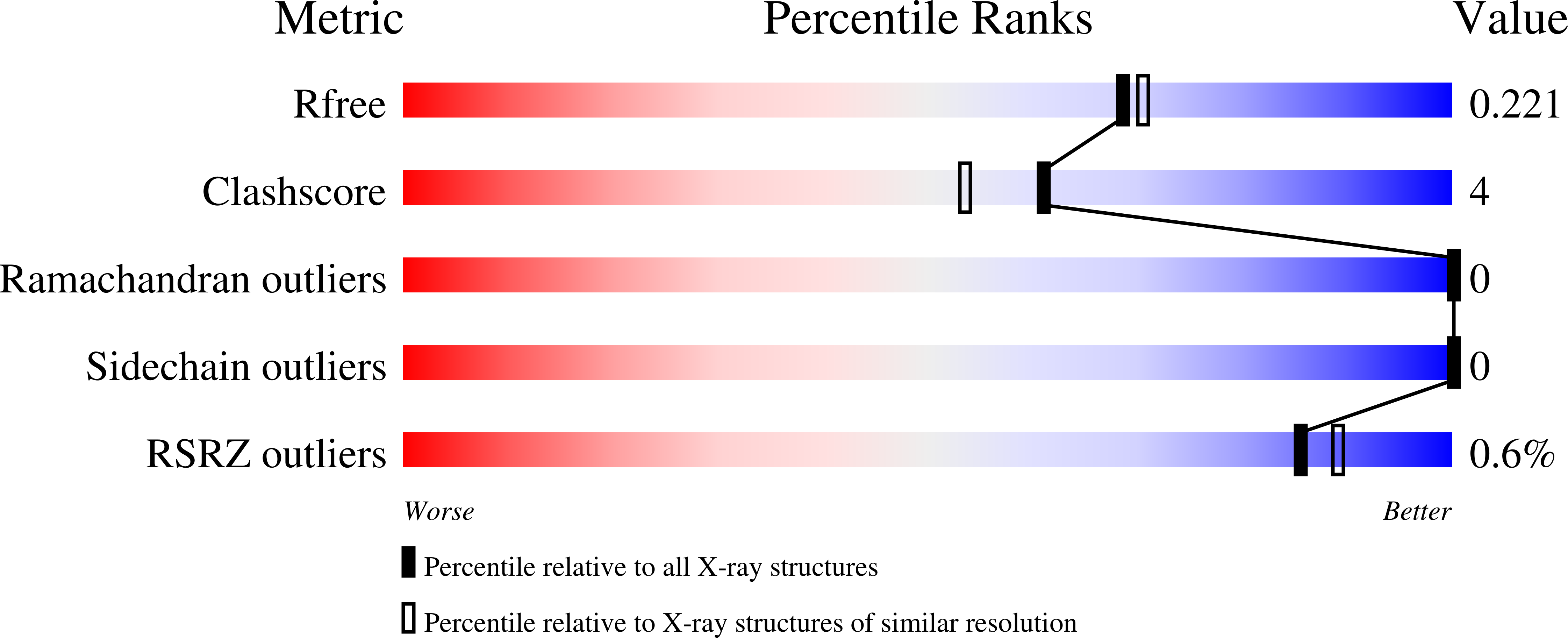
1.95 Å
R-Value Free:
0.22
R-Value Work:
0.17
R-Value Observed:
0.18
Space Group:
P 21 2 21
Macromolecular Entities
Polymer Type:polypeptide(L)
Molecule:Phosphoribosyl isomerase A
Gene (Uniprot):priA
Chain IDs:A, B
Chain Length:244
Number of Molecules:2
Biological Source:Mycobacterium tuberculosis
Ligand Molecules
Primary Citation
Crystal structure of the bifunctional enzyme phosphoribosyl isomerase A from Mycobacterium tuberculosis with bound light-controllable inhibitor.
Acta Crystallogr.,Sect.F 82 385 390 (2026)
PMID: 42861558 DOI: 10.1107/S2053230X26009684

Abstact

The enzyme phosphoribosyl isomerase A from Mycobacterium tuberculosis (mtPriA) catalyzes two related reactions of bi- and monophosphorylated substrates within the histidine and tryptophan biosynthetic pathways. We have determined the crystal structure of mtPriA with a bound light-controllable 1,2-dithienylethene inhibitor (DTE LC6) at a resolution of 1.95 A. DTE LC6 contains two terminal phosphate groups, and we found one inhibitor molecule (DTE LC6-1) simultaneously bound to the two known functional phosphate-binding sites of mtPriA. Unexpectedly, another inhibitor molecule (DTE LC6-2) was shown to bind to a hitherto unidentified third phosphate-binding site and thus bridges two crystallographic symmetry-related mtPriA proteins. Our study explains the previously reported inhibitory effect of DTE LC6 on the catalytic activity of mtPriA and raises the question of the significance of the third phosphate-binding site in mtPriA.

Legend

Protein

Chemical

Disease

Primary Citation of related structures
Feedback Form
Name
Email
Institute
Feedback