

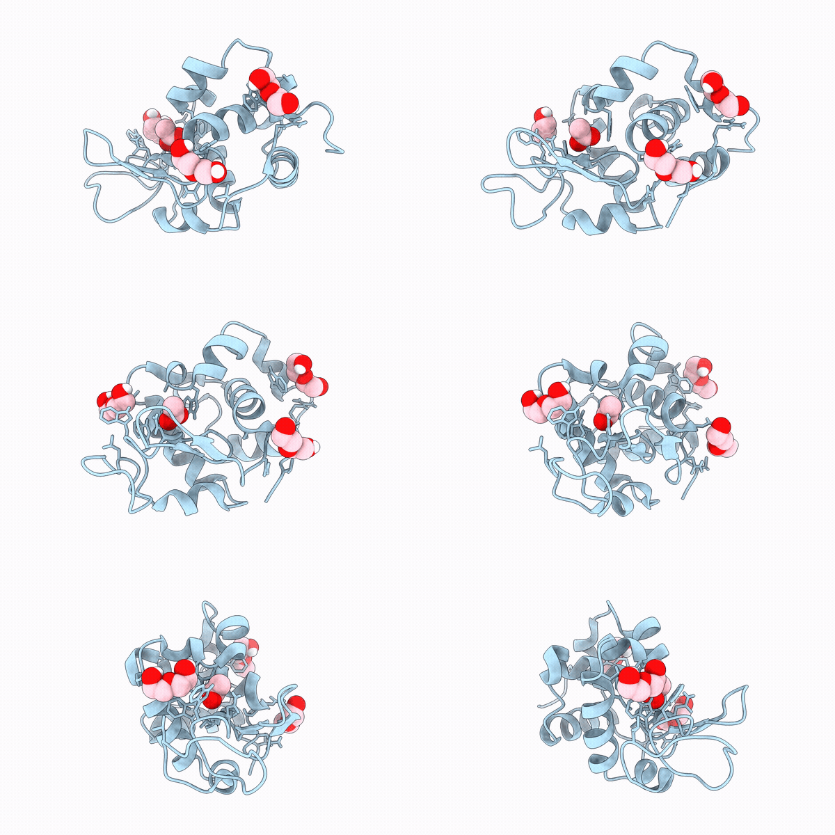
30DB image
Deposition Date 2026-04-21
Release Date 2026-10-28
Last Version Date 2026-10-28
Entry Detail
PDB ID:
30DB
Keywords:
Title:
Hen egg white lysozyme complexed with PEG, ACT, and CL (in meso crystallization)
Biological Source:
Source Organism(s):
Gallus gallus (Taxon ID: 9031)
Method Details:
Experimental Method:
Resolution:
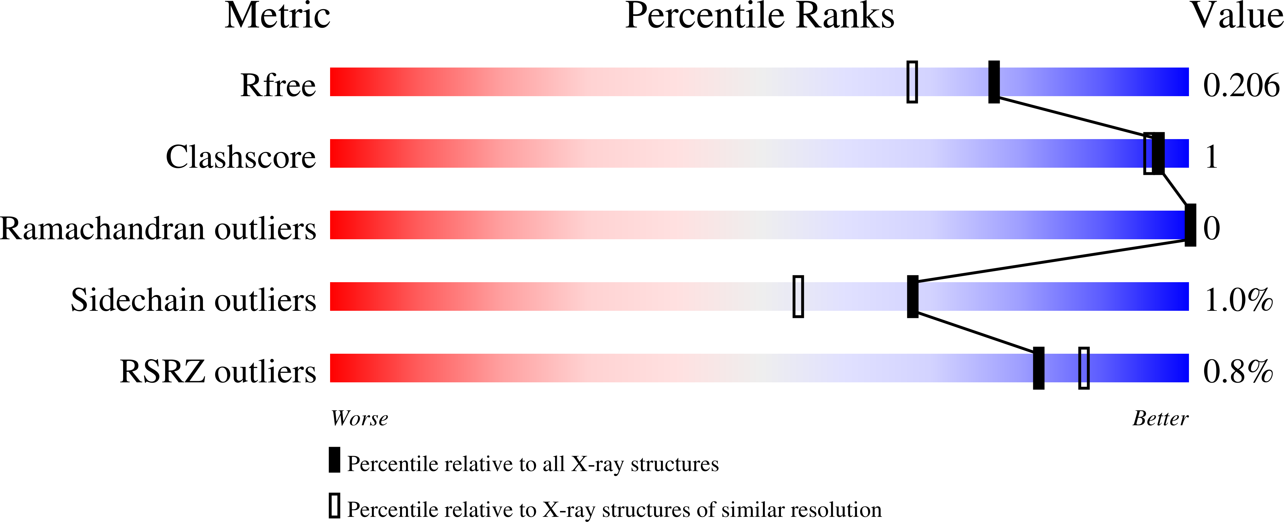
1.74 Å
R-Value Free:
0.20
R-Value Work:
0.17
R-Value Observed:
0.17
Space Group:
P 43 21 2
Macromolecular Entities
Polymer Type:polypeptide(L)
Molecule:Lysozyme C
Gene (Uniprot):LYZ
Chain IDs:A
Chain Length:129
Number of Molecules:1
Biological Source:Gallus gallus
Primary Citation
A low-cost, portable polarizing digital light microscope for monitoring crystal growth and for crystal harvesting.
J.Appl.Crystallogr. 59 1693 1706 (2026)
PMID: 42835515 DOI: 10.1107/S1600576726007880

Abstact

Motivated by the need for a convenient way to demonstrate the growth of crystals in glass plates by the lipid cubic phase method at crystallization workshops and courses, we developed a lightweight, portable, compact and inexpensive digital microscope device. The device is built around hand-held Wi-Fi and hardwired digital microscopes that are available commercially at low cost and that can use cell phones and personal computers for imaging without the need for unwieldy eyepieces. The device provides illumination from above and below the crystallization plate over a range of brightness levels. It can be used with regular white light and with polarized light to detect birefringent crystals. While not the focus of this study, the new device has been shown to also work with batch and vapour-diffusion plates. Remote monitoring of crystallization plates in a cold cabinet or walk-in refrigerator at 4 degrees C is possible with the Wi-Fi microscope. Crystals grown in lipid cubic phase and vapour-diffusion crystallization plates were harvested with the aid of the new device and used for diffraction data collection, leading to structure determination at high resolution. Most parts of the device were 3D printed in polylactic acid plastic. The corresponding STL files, included as part of this publication, can be used to make replicates of the current microscope and to produce bespoke devices suited to the specific needs of the user. We envision using the new device for outreach activities at primary, secondary and third-level schools, and at science fairs in support of the citizen scientist. Relatedly, in this study examples are included of recorded images of plant and animal tissue sections. One reveals birefringent materials in a tomato leaf. Another shows optically active crystals of calcium oxalate in onion tunic tissue.

Legend

Protein

Chemical

Disease

Primary Citation of related structures
Feedback Form
Name
Email
Institute
Feedback