Abstact
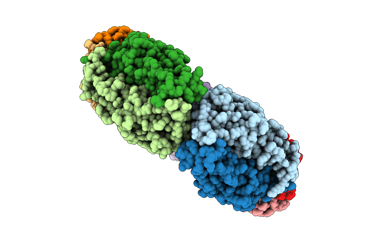
Rat heme-binding protein 23 (HBP23)/peroxiredoxin (Prx I) belongs to the 2-Cys peroxiredoxin type I family and exhibits peroxidase activity coupled with reduced thioredoxin (Trx) as an electron donor. We analyzed the dimer-oligomer interconversion of wild-type and mutant HBP23/Prx I by gel filtration and found that the C52S and C173S mutants existed mostly as decamers, whereas the wild type was a mixture of various forms, favoring the decamer at higher protein concentration and lower ionic salt concentration and in the presence of dithiothreitol. The C83S mutant was predominantly dimeric, in agreement with a previous crystallographic analysis (Hirotsu, S., Abe, Y., Okada, K., Nagahara, N., Hori, H., Nishino, T., and Hakoshima, T. (1999) Proc. Natl. Acad. Sci. U. S. A. 96, 12333-12338). X-ray diffraction analysis of the decameric C52S mutant revealed a toroidal structure (diameter, approximately 130A; inside diameter, approximately 55A; thickness, approximately 45A). In contrast to human Prx I, which was recently reported to exist predominantly as the decamer with Cys(83)-Cys(83) disulfide bonds at all dimer-dimer interfaces, rat HBP23/Prx I has a Cys(83)-Cys(83) disulfide bond at only one dimer-dimer interface (S-S separation of approximately 2.1A), whereas the interactions at the other interfaces (mean S-S separation of 3.6A) appear to involve hydrophobic and van der Waals forces. This finding is consistent with gel filtration analyses showing that the protein readily interconverts between dimer and oligomeric forms. The C83S mutant exhibited similar peroxidase activity to the wild type, which is exclusively dimeric, in the Trx/Trx reductase system. At higher concentrations, where the protein was mostly decameric, less efficient attack of reduced Trx was observed in a [(14)C]iodoacetamide incorporation experiment. We suggest that the dimerdecamer interconversion may have a regulatory role.