

26HG image
Deposition Date 2026-04-30
Release Date 2026-10-28
Last Version Date 2026-11-11
Entry Detail
PDB ID:
26HG
Keywords:
Title:
X-ray crystal structure of the holo-form of diflavin-linked disulfide oxidoreductase
Biological Source:
Source Organism(s):
Expression System(s):
Method Details:
Experimental Method:
Resolution:
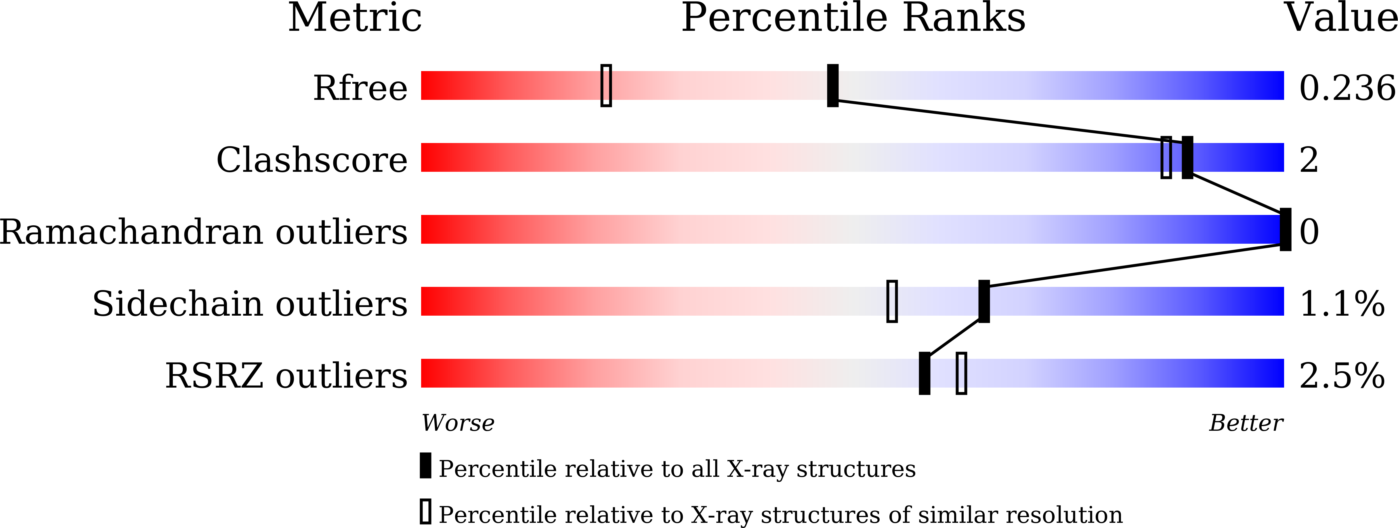
1.70 Å
R-Value Free:
0.23
R-Value Work:
0.20
R-Value Observed:
0.20
Space Group:
C 1 2 1
Macromolecular Entities
Polymer Type:polypeptide(L)
Molecule:Slr0600 protein
Gene (Uniprot):slr0600
Chain IDs:A
Chain Length:326
Number of Molecules:1
Biological Source:Synechocystis sp. PCC 6803
Ligand Molecules
Primary Citation
Characterisation of FAD binding to the apo-form of diflavin-linked disulfide oxidoreductase derived from cyanobacterium Synechocystis sp. PCC6803.
Biochem.J. 483 2227 2240 (2026)
PMID: 42821309 DOI: 10.1042/BCJ20260320

Abstact

Diflavin-linked disulfide oxidoreductase (DDOR) from the cyanobacterium Synechocystis sp. PCC6803 is an NADPH-thioredoxin reductase (NTR) superfamily enzyme that forms a homodimer containing two FAD molecules (FAD1 and FAD2) per monomer. FAD1 is conserved in the NTR superfamily, while FAD2 is characteristic of DDOR. Under aerobic conditions, thiol reduction of DDOR produces an apparently air-stable neutral blue semiquinone, but the respective roles of FAD1 and FAD2 in the reduction process remain unclear. To address this, spectroscopic, structural, and mutational analyses were performed to investigate the contributions of the two FAD molecules. Recombinant apo- and holo-form of DDOR (holo-DDOR) synthesised in Escherichia coli cells were separated and purified. Fluorescence titration of the apo-form of DDOR (apo-DDOR) with FAD revealed rapid binding of two FADs. Stopped-flow analysis indicated biphasic binding of FAD to apo-DDOR. X-ray crystal structures of apo- and holo-DDORs exhibited pronounced conformational differences in the FAD-binding sites, suggesting that the first FAD may bind to the FAD1-binding site and subsequently induce formation of the FAD2-binding site. Systematic replacement of amino acid residues in the FAD2-binding site showed that mutations at the characteristic loop region bearing the GKKDSLY motif resulted in binding of three flavin species of FAD, flavin mononucleotide, and riboflavin. Moreover, replacement of both Trp53 and Tyr143 at the binding site of the isoalloxazine ring of FAD2 with Ala residues yielded exclusively the apo-form. These results indicate that the loop region restricts the flavin species bound at the FAD2-binding site, and Trp53 and Tyr143 are important for FAD bindings to both the FAD1- and FAD2-binding sites.

Legend

Protein

Chemical

Disease

Primary Citation of related structures
Feedback Form
Name
Email
Institute
Feedback