1W6S image
Deposition Date 2004-08-23
Release Date 2004-12-21
Last Version Date 2024-11-13
Entry Detail
PDB ID:
1W6S
Keywords:
Title:
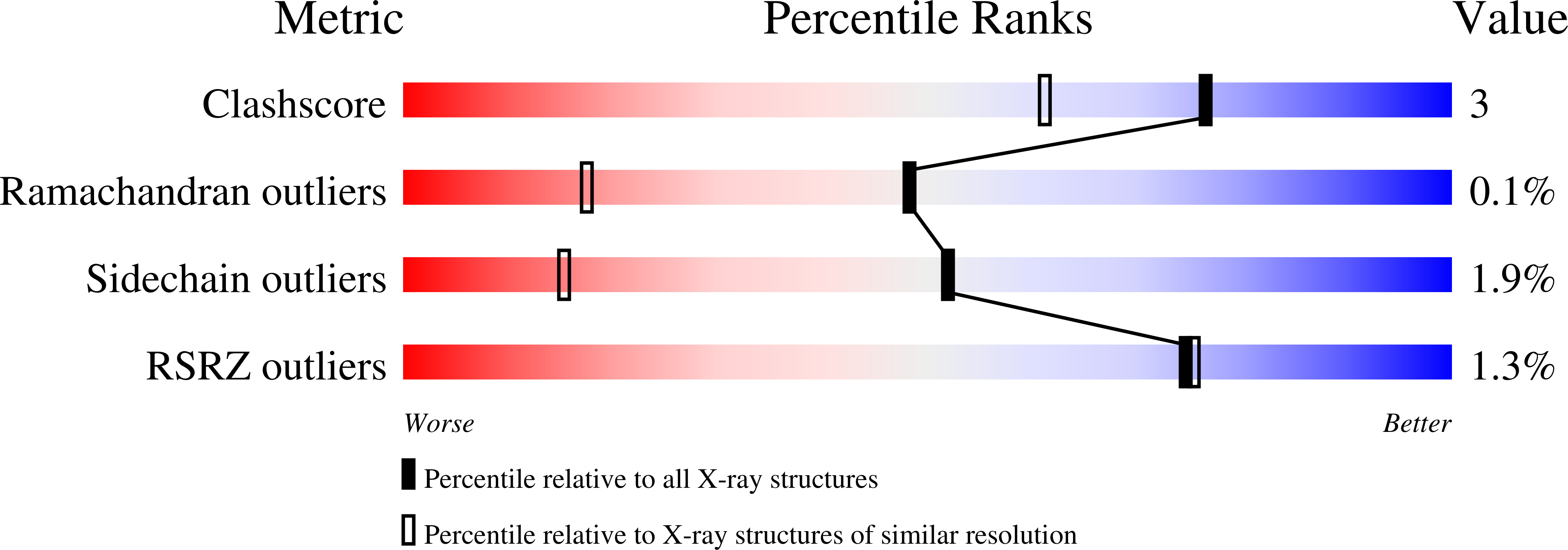
The high resolution structure of methanol dehydrogenase from methylobacterium extorquens
Biological Source:
Source Organism(s):
Method Details:
Experimental Method:
Resolution:
1.20 Å
R-Value Free:
0.17
R-Value Observed:
0.15
Space Group:
P 1
Macromolecular Entities
Structures with similar UniProt ID
Protein Blast
Polymer Type:polypeptide(L)
Molecule:METHANOL DEHYDROGENASE SUBUNI
Gene (Uniprot):moxF
Chain IDs:A, C
Chain Length:599
Number of Molecules:2
Biological Source:METHYLOBACTERIUM EXTORQUENS
Structures with similar UniProt ID
Protein Blast
Polymer Type:polypeptide(L)
Molecule:METHANOL DEHYDROGENASE SUBUNI
Gene (Uniprot):moxI
Chain IDs:B, D
Chain Length:74
Number of Molecules:2
Biological Source:METHYLOBACTERIUM EXTORQUENS
Primary Citation
The Atomic Resolution Structure of Methanol Dehydrogenase from Methylobacterium Extorquens
Acta Crystallogr. D Biol. Crystallogr. 61 75 ? (2005)
PMID: 15608378 DOI: 10.1107/S0907444904026964

Abstact

The crystal structure of methanol dehydrogenase (MDH) from Methylobacterium extorquens has been refined without stereochemical restraints at a resolution of 1.2 A. The high-resolution data have defined the conformation of the tricyclic pyrroloquinoline quinone (PQQ) cofactor ring as entirely planar. The detailed definition of the active-site geometry has shown many features that are similar to the quinohaemo-protein alcohol dehydrogenases from Comamonas testosteroni and Pseudomonas putida, both of which possess MDH-like and cytochrome c-like domains. Conserved features between the two types of PQQ-containing enzyme suggest a common pathway for electron transfer between MDH and its physiological electron acceptor cytochrome cL. A pathway for proton transfer from the active site to the bulk solvent is also suggested.

Legend

Protein

Chemical

Disease

Primary Citation of related structures
Feedback Form
Name
Email
Institute
Feedback