Deposition Date
2001-05-11
Release Date
2001-08-23
Last Version Date
2024-11-13
Entry Detail
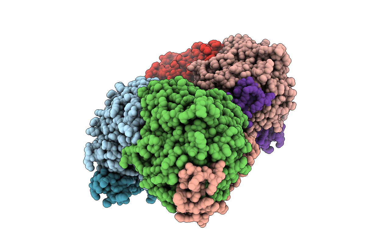
PDB ID:
1H4J
Keywords:
Title:
Methylobacterium extorquens methanol dehydrogenase D303E mutant
Biological Source:
Source Organism(s):
Methylobacterium extorquens (Taxon ID: 408)
Expression System(s):
Method Details:
Experimental Method:
Resolution:
3.00 Å
R-Value Free:
0.21
R-Value Work:
0.18
R-Value Observed:
0.18
Space Group:
P 1 21 1