

1BYD image
Deposition Date 1994-01-25
Release Date 1994-07-31
Last Version Date 2024-02-07
Entry Detail
PDB ID:
1BYD
Title:
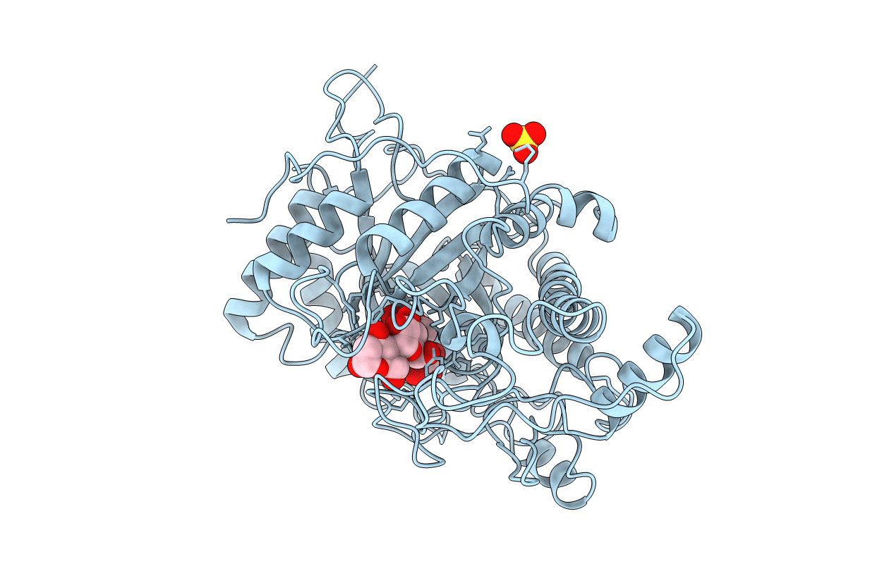
CRYSTAL STRUCTURES OF SOYBEAN BETA-AMYLASE REACTED WITH BETA-MALTOSE AND MALTAL: ACTIVE SITE COMPONENTS AND THEIR APPARENT ROLE IN CATALYSIS
Biological Source:
Source Organism(s):
Glycine max (Taxon ID: 3847)
Method Details:
Experimental Method:
Resolution:
2.20 Å
R-Value Work:
0.14
R-Value Observed:
0.14
Space Group:
P 31 2 1
Macromolecular Entities
Polymer Type:polypeptide(L)
Molecule:BETA-AMYLASE
Gene (Uniprot):BMY1
Chain IDs:A
Chain Length:495
Number of Molecules:1
Biological Source:Glycine max
Ligand Molecules
Peptide-like Molecules
PRD_900048
Primary Citation
Crystal structures of soybean beta-amylase reacted with beta-maltose and maltal: active site components and their apparent roles in catalysis.
Biochemistry 33 7779 7787 (1994)
PMID: 8011643 DOI: 10.1021/bi00191a005

Abstact

The crystal structures of catalytically competent soybean beta-amylase, unliganded and bathed with small substrates (beta-maltose, maltal), were determined at 1.9-2.2-A resolution. Two molecules of beta-maltose substrate bind to the protein in tandem, with some maltotetraose enzymic condensation product sharing the same binding sites. The beta-amylase soaked with maltal shows a similar arrangement of two bound molecules of 2-deoxymaltose, the enzymic hydration product. In each case the nonreducing ends of the saccharide ligands are oriented toward the base of the protein's active site pocket. The catalytic center, located between the bound disaccharides and found deeper in the pocket than where the inhibitor alpha-cyclodextrin binds, is characterized by the presence of oppositely disposed carboxyl groups of two conserved glutamic acid residues. The OE2 carboxyl of Glu 186 is below the plane of the penultimate glucose residue (Glc 2) of bound maltotetraose, 2.6 A from the oxygen atom of that ligand's penultimate alpha-1,4-glucosidic linkage. The OE2 carboxyl of Glu 380 lies above the plane of Glc 2, 2.8 A from the O-1 atom of the more deeply bound beta-maltose. Saccharide binding does not alter the spatial coordinates of these two carboxyl groups or the overall conformation of the 57-kDa protein. However, the saccharide complexes of the active enzyme are associated with a significant (10 A) local conformational change in a peptide segment of a loop (L3) that borders the active site pocket.(ABSTRACT TRUNCATED AT 250 WORDS)

Legend

Protein

Chemical

Disease

Primary Citation of related structures
Feedback Form
Name
Email
Institute
Feedback