

Search Count: 31
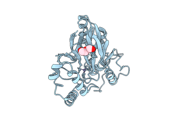 |
Crystal Structure Of The Persulfide Dioxygenase (Pdo - Pa2915) From Pseudomonas Aeruginosa
Organism: Pseudomonas aeruginosa
Method: X-RAY DIFFRACTION Resolution:2.06 Å Release Date: 2025-08-06 Classification: OXIDOREDUCTASE Ligands: PEG, ZN, CL |
Organism: Pseudomonas aeruginosa
Method: X-RAY DIFFRACTION
Release Date: 2025-08-06
Ligands: PEG, ZN, CL
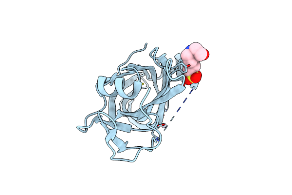 |
Crystal Structure Of Protein D: Defoliating Toxin Form Fusarium Oxysporum F.Sp. Vasinfectum
Organism: Fusarium oxysporum
Method: X-RAY DIFFRACTION Resolution:1.47 Å Release Date: 2025-05-28 Classification: TOXIN Ligands: EPE, HG |
Organism: Fusarium oxysporum
Method: X-RAY DIFFRACTION
Release Date: 2025-05-28
Ligands: EPE, HG
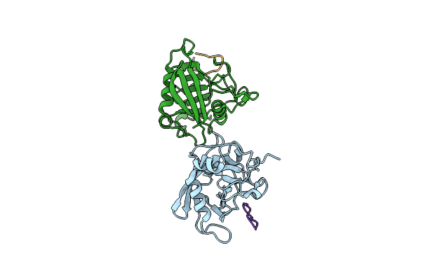 |
Crystal Structure Of Cyclophilin Tgcyp23 From Toxoplasma Gondii In Complex With Nim811 (N-Methyl-4-Isoleucine Cyclosporin)
Organism: Toxoplasma gondii, Synthetic construct
Method: X-RAY DIFFRACTION Resolution:1.17 Å Release Date: 2024-10-30 Classification: ISOMERASE |
Organism: Toxoplasma gondii, Synthetic construct
Method: X-RAY DIFFRACTION
Release Date: 2024-10-30
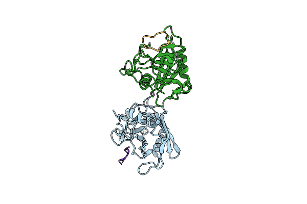 |
Crystal Structure Of Cyclophilin Tgcyp23 From Toxoplasma Gondii In Complex With Alisporivir (Nonimmunosuppressive Analogue Of Cyclosporin)
Organism: Toxoplasma gondii, Synthetic construct
Method: X-RAY DIFFRACTION Resolution:1.20 Å Release Date: 2024-10-30 Classification: ISOMERASE |
Organism: Toxoplasma gondii, Synthetic construct
Method: X-RAY DIFFRACTION
Release Date: 2024-10-30
 |
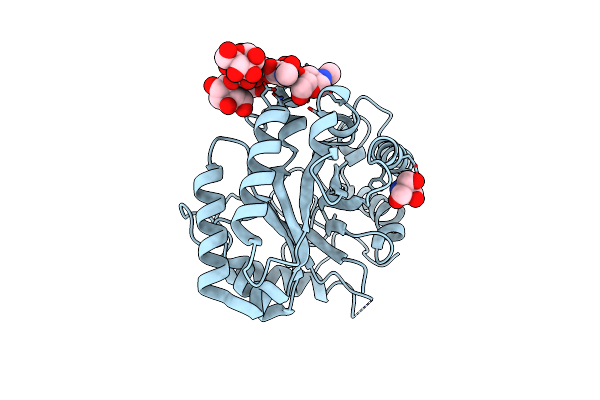
Crystal Structure Of Cyclophilin Tgcyp23 From Toxoplasma Gondii In Complex With Dihydro Cyclosporin A
Organism: Toxoplasma gondii, Synthetic construct
Method: X-RAY DIFFRACTION Resolution:1.20 Å Release Date: 2024-10-30 Classification: ISOMERASE |
Organism: Toxoplasma gondii, Synthetic construct
Method: X-RAY DIFFRACTION
Release Date: 2024-10-30
 |
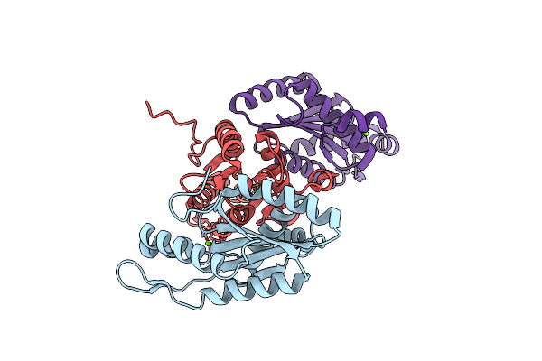
Crystal Structure Of The Catalytic Domain Of G7048 From Penicillium Sumatraense
Organism: Penicillium sumatraense
Method: X-RAY DIFFRACTION Resolution:1.90 Å Release Date: 2022-12-28 Classification: HYDROLASE Ligands: TRS |
Organism: Penicillium sumatraense
Method: X-RAY DIFFRACTION
Release Date: 2022-12-28
Ligands: TRS
 |
Structure Of A Delta-N Mutant - E232Start - Of Pa1120 (Tpbb Or Yfin) From Pseudomonas Aeruginosa (Pao1) Comprising Only The Ggdef Domain
Organism: Pseudomonas aeruginosa pao1
Method: X-RAY DIFFRACTION Resolution:2.80 Å Release Date: 2021-07-07 Classification: LIGASE Ligands: MG |
Organism: Pseudomonas aeruginosa pao1
Method: X-RAY DIFFRACTION
Release Date: 2021-07-07
Ligands: MG
 |
Thioredoxin Glutathione Reductase From Schistosoma Mansoni In Complex With Indole-3-Carbinol
Organism: Schistosoma mansoni
Method: X-RAY DIFFRACTION Resolution:1.90 Å Release Date: 2021-05-19 Classification: PROTEIN BINDING Ligands: FAD, FXK, DMS, PGE, K |
Organism: Schistosoma mansoni
Method: X-RAY DIFFRACTION
Release Date: 2021-05-19
Ligands: FAD, FXK, DMS, PGE, K
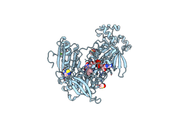 |
Thioredoxin Glutathione Reductase From Schistosoma Mansoni In Complex With 4-Aminopiazthiole
Organism: Schistosoma mansoni
Method: X-RAY DIFFRACTION Resolution:2.15 Å Release Date: 2021-05-19 Classification: PROTEIN BINDING Ligands: FAD, DMS, BTE, CA |
Organism: Schistosoma mansoni
Method: X-RAY DIFFRACTION
Release Date: 2021-05-19
Ligands: FAD, DMS, BTE, CA
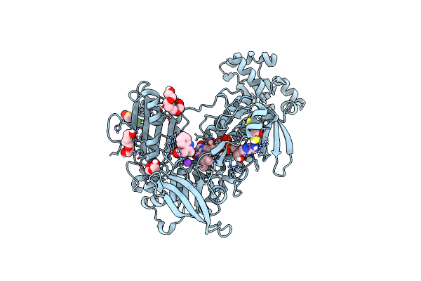 |
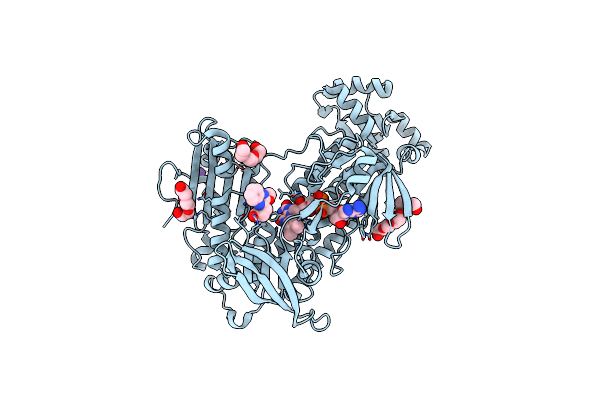
Thioredoxin Glutathione Reductase From Schistosoma Mansoni In Complex With 2-Methylindole-3-Acetic Acid
Organism: Schistosoma mansoni
Method: X-RAY DIFFRACTION Resolution:1.80 Å Release Date: 2021-05-19 Classification: PROTEIN BINDING Ligands: FAD, K, PG4, DMS, QN5, CA, PEG, DTT, PGE |
Organism: Schistosoma mansoni
Method: X-RAY DIFFRACTION
Release Date: 2021-05-19
Ligands: FAD, K, PG4, DMS, QN5, CA, PEG, DTT, PGE
 |
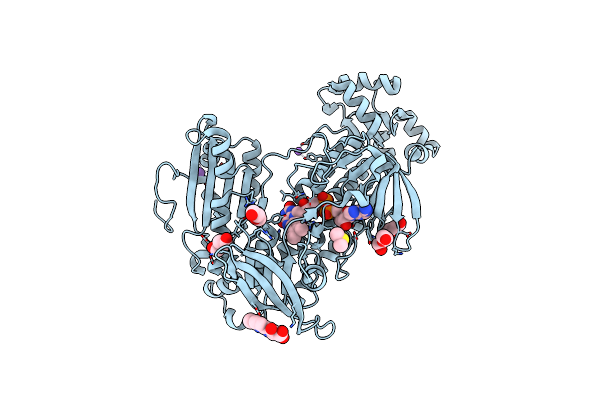
Thioredoxin Glutathione Reductase From Schistosoma Mansoni In Complex With 3-(3-Methoxyquinoxalin-2-Yl)Propanoic Acid
Organism: Schistosoma mansoni
Method: X-RAY DIFFRACTION Resolution:1.70 Å Release Date: 2021-05-19 Classification: PROTEIN BINDING Ligands: FAD, K, PEG, PGE, EY7 |
Organism: Schistosoma mansoni
Method: X-RAY DIFFRACTION
Release Date: 2021-05-19
Ligands: FAD, K, PEG, PGE, EY7
 |
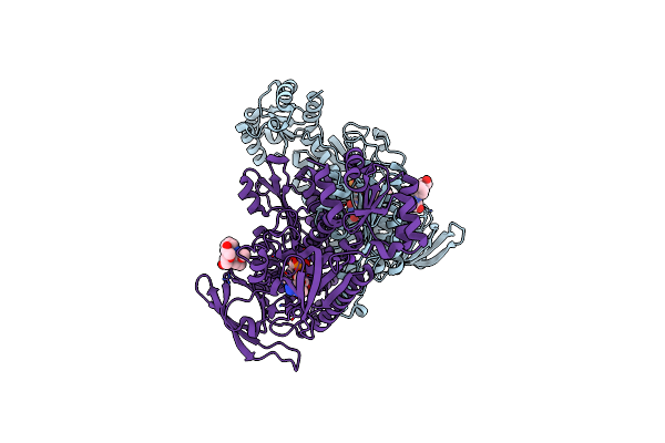
Thioredoxin Glutathione Reductase From Schistosoma Mansoni In Complex With 4-Hydroxy-7-Methyl-1,8-Naphthyridine-3-Carboxylic Acid
Organism: Schistosoma mansoni
Method: X-RAY DIFFRACTION Resolution:1.45 Å Release Date: 2021-05-19 Classification: PROTEIN BINDING Ligands: FAD, PEG, DMS, SJN, K, CL, NA |
Organism: Schistosoma mansoni
Method: X-RAY DIFFRACTION
Release Date: 2021-05-19
Ligands: FAD, PEG, DMS, SJN, K, CL, NA
 |
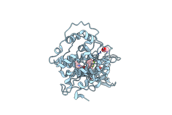
Thioredoxin Glutathione Reductase From Schistosoma Mansoni In Complex With 3-(3-Methoxyquinoxalin-2-Yl)Propanoic Acid At 24 Hours Of Soaking
Organism: Schistosoma mansoni
Method: X-RAY DIFFRACTION Resolution:2.70 Å Release Date: 2021-05-19 Classification: PROTEIN BINDING Ligands: FAD, EY7, CL, PEG, K |
Organism: Schistosoma mansoni
Method: X-RAY DIFFRACTION
Release Date: 2021-05-19
Ligands: FAD, EY7, CL, PEG, K
 |
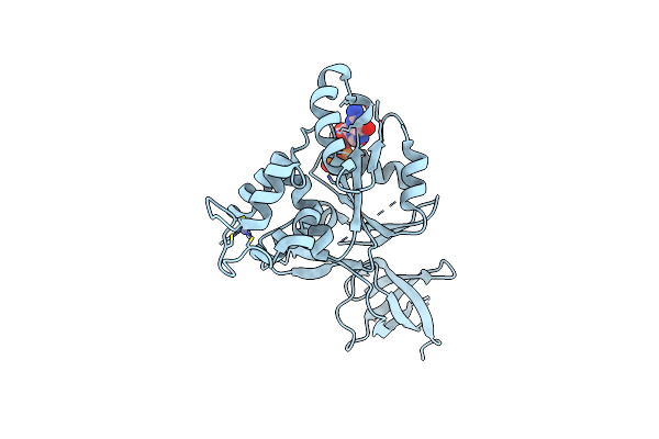
Glycogen Synthase Kinase-3 Beta (Gsk3B) In Complex With Ligand 1
Organism: Homo sapiens
Method: X-RAY DIFFRACTION Resolution:2.14 Å Release Date: 2020-09-16 Classification: TRANSFERASE Ligands: N1Q, ACT |
Organism: Homo sapiens
Method: X-RAY DIFFRACTION
Release Date: 2020-09-16
Ligands: N1Q, ACT
 |
Crystal Structure Of Rsga From Pseudomonas Aeruginosa
Organism: Pseudomonas aeruginosa pao1
Method: X-RAY DIFFRACTION Resolution:2.90 Å Release Date: 2019-06-26 Classification: RNA BINDING PROTEIN Ligands: ZN, GDP |
Organism: Pseudomonas aeruginosa pao1
Method: X-RAY DIFFRACTION
Release Date: 2019-06-26
Ligands: ZN, GDP
 |
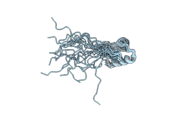
Xi Class Gst From Natrialba Magadii
Organism: natrialba magadii atcc 43099
Method: X-RAY DIFFRACTION Resolution:2.61 Å Release Date: 2019-02-13 Classification: TRANSFERASE Ligands: SO4 |
Organism: natrialba magadii atcc 43099
Method: X-RAY DIFFRACTION
Release Date: 2019-02-13
Ligands: SO4
 |
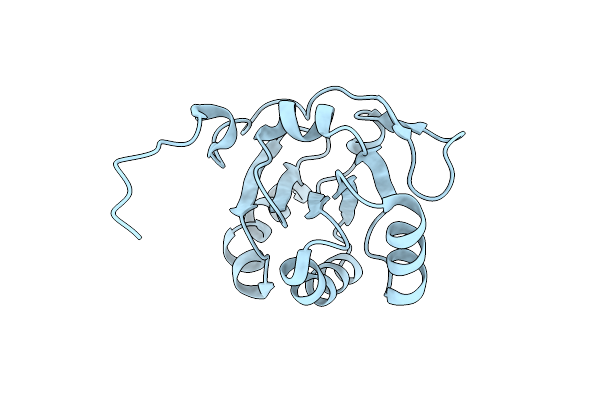
Nmr Structure Of Npm1_C70
Organism: Homo sapiens
Method: SOLUTION NMR Release Date: 2012-06-27 Classification: DNA BINDING PROTEIN, CHAPERONE |
Organism: Homo sapiens
Method: SOLUTION NMR
Release Date: 2012-06-27
 |
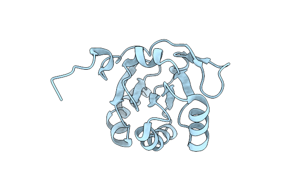
Crystal Structure Of The Periplasmic Soluble Domain Of Reduced Ccmg From Pseudomonas Aeruginosa
Organism: Pseudomonas aeruginosa
Method: X-RAY DIFFRACTION Resolution:1.75 Å Release Date: 2010-04-07 Classification: OXIDOREDUCTASE |
Organism: Pseudomonas aeruginosa
Method: X-RAY DIFFRACTION
Release Date: 2010-04-07
 |
Crystal Structure Of The Periplasmic Soluble Domain Of Oxidized Ccmg From Pseudomonas Aeruginosa
Organism: Pseudomonas aeruginosa
Method: X-RAY DIFFRACTION Resolution:2.20 Å Release Date: 2010-04-07 Classification: OXIDOREDUCTASE |
Organism: Pseudomonas aeruginosa
Method: X-RAY DIFFRACTION
Release Date: 2010-04-07
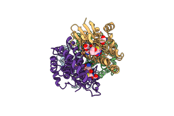 |
Crystal Structure Of Mu Class Glutathione S-Transferase (Gstm2-2) In Complex With Glutathione And 6-(7-Nitro-2,1,3-Benzoxadiazol-4-Ylthio)Hexanol (Nbdhex)
Organism: Homo sapiens
Method: X-RAY DIFFRACTION Resolution:2.50 Å Release Date: 2009-10-27 Classification: TRANSFERASE/TRANSFERASE INHIBITOR Ligands: BYG, GSH, EDO |
Organism: Homo sapiens
Method: X-RAY DIFFRACTION
Release Date: 2009-10-27
Ligands: BYG, GSH, EDO