

Search Count: 8,574
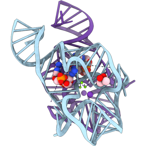 |
Crystal Structure Of The C-Di-Gmp-Ii Riboswitch C-Di-Gmp-Ii-Bh Bound With 2',3'-Cgamp
Organism: Halalkalibacterium halodurans c-125
Method: X-RAY DIFFRACTION Resolution:2.15 Å Release Date: 2026-10-21 Classification: RNA Ligands: 1SY, MG, K, CAC |
Organism: Halalkalibacterium halodurans c-125
Method: X-RAY DIFFRACTION
Release Date: 2026-10-21
Ligands: 1SY, MG, K, CAC
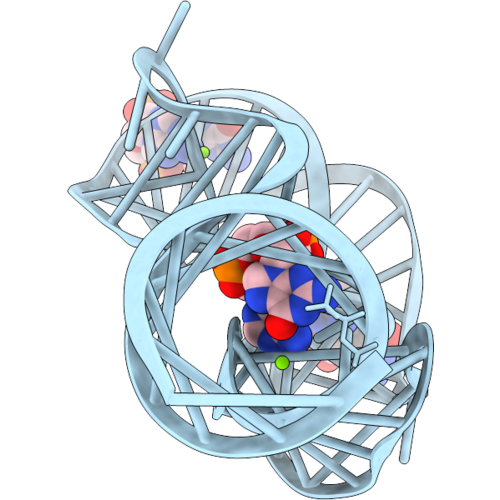 |
Crystal Structure Of A Variant C-Di-Gmp-Ii Riboswitch C-Di-Gmp-Ii-Bh_A9G/U55C Bound With 3',3'-Cgamp
Organism: Halalkalibacterium halodurans c-125
Method: X-RAY DIFFRACTION Resolution:2.04 Å Release Date: 2026-10-21 Classification: RNA Ligands: 4BW, MG |
Organism: Halalkalibacterium halodurans c-125
Method: X-RAY DIFFRACTION
Release Date: 2026-10-21
Ligands: 4BW, MG
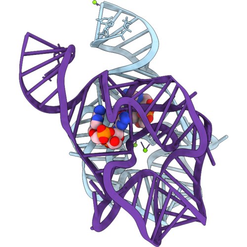 |
Crystal Structure Of A Variant C-Di-Gmp-Ii Riboswitch C-Di-Gmp-Ii-Bh_A9G/U55C Bound With 2',3'-Cgamp
Organism: Halalkalibacterium halodurans c-125
Method: X-RAY DIFFRACTION Resolution:2.90 Å Release Date: 2026-10-21 Classification: RNA Ligands: 1SY, MG |
Organism: Halalkalibacterium halodurans c-125
Method: X-RAY DIFFRACTION
Release Date: 2026-10-21
Ligands: 1SY, MG
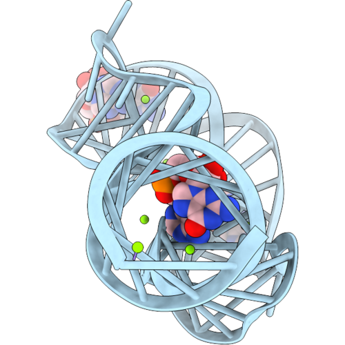 |
Crystal Structure Of A Variant C-Di-Gmp-Ii Riboswitch C-Di-Gmp-Ii-Bh Bound With 3',3'-Cgamp
Organism: Halalkalibacterium halodurans c-125
Method: X-RAY DIFFRACTION Resolution:1.70 Å Release Date: 2026-10-21 Classification: RNA Ligands: 4BW, MG |
Organism: Halalkalibacterium halodurans c-125
Method: X-RAY DIFFRACTION
Release Date: 2026-10-21
Ligands: 4BW, MG
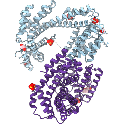 |
Protein With 8.3 Knot
Organism: Lachnospiraceae bacterium
Method: X-RAY DIFFRACTION Resolution:2.54 Å Release Date: 2026-10-21 Classification: UNKNOWN FUNCTION Ligands: EDO, SO4, PEG |
Organism: Lachnospiraceae bacterium
Method: X-RAY DIFFRACTION
Release Date: 2026-10-21
Ligands: EDO, SO4, PEG
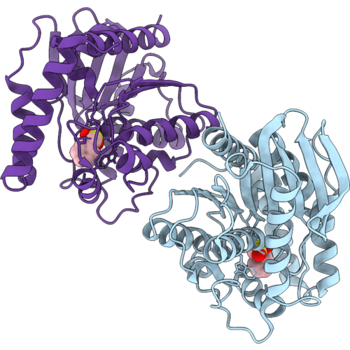 |
Actinomycetes Hydrolase Inhibited By Pmsf
Organism: Actinomycetes bacterium
Method: X-RAY DIFFRACTION Resolution:1.60 Å Release Date: 2026-10-21 Classification: HYDROLASE Ligands: PMS |
Organism: Actinomycetes bacterium
Method: X-RAY DIFFRACTION
Release Date: 2026-10-21
Ligands: PMS
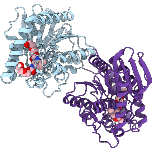 |
Actinomycetes Hydrolase (S148A) Bound To 2,4-Tda-Deg
Organism: Actinomycetes bacterium
Method: X-RAY DIFFRACTION Resolution:1.15 Å Release Date: 2026-10-21 Classification: HYDROLASE Ligands: A1KUO |
Organism: Actinomycetes bacterium
Method: X-RAY DIFFRACTION
Release Date: 2026-10-21
Ligands: A1KUO
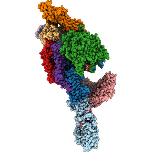 |
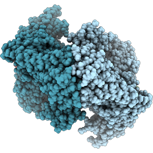
Lpt Translocon Complex From Flavobacter Johnsoniae Built In Composite Volume
Organism: Flavobacterium johnsoniae uw101
Method: ELECTRON MICROSCOPY Release Date: 2026-10-21 Classification: MEMBRANE PROTEIN Ligands: GLA |
Organism: Flavobacterium johnsoniae uw101
Method: ELECTRON MICROSCOPY
Release Date: 2026-10-21
Ligands: GLA
 |
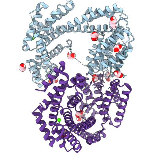
Mycobacterium Tuberculosis Gyrase Holocomplex With 150 Bp Dna And Corramycin
Organism: Mycobacterium tuberculosis, Escherichia coli, Corallococcus coralloides
Method: ELECTRON MICROSCOPY Release Date: 2026-10-21 Classification: DNA BINDING PROTEIN |
Organism: Mycobacterium tuberculosis, Escherichia coli, Corallococcus coralloides
Method: ELECTRON MICROSCOPY
Release Date: 2026-10-21
 |
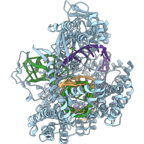
Protein With Novel 8.3 Knotted Fold - Eight Fold (Extraordinarily Intricate Globular Helical Tangled Fold).
Organism: Lachnospiraceae bacterium
Method: X-RAY DIFFRACTION Resolution:2.92 Å Release Date: 2026-10-21 Classification: UNKNOWN FUNCTION Ligands: EDO, PEG, GOL, CA, ACT |
Organism: Lachnospiraceae bacterium
Method: X-RAY DIFFRACTION
Release Date: 2026-10-21
Ligands: EDO, PEG, GOL, CA, ACT
 |
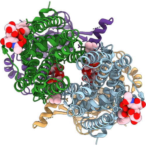
Cryo-Em Structure Of The Lbcas12A-Lid2-Crrna-Dna Complex
Organism: Lachnospiraceae bacterium nd2006
Method: ELECTRON MICROSCOPY Release Date: 2026-10-14 Classification: RNA BINDING PROTEIN Ligands: MG |
Organism: Lachnospiraceae bacterium nd2006
Method: ELECTRON MICROSCOPY
Release Date: 2026-10-14
Ligands: MG
 |
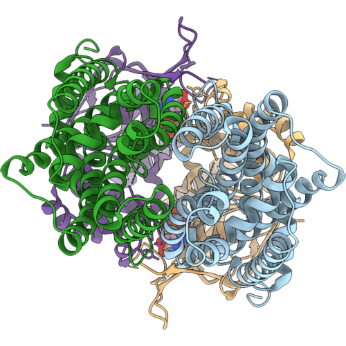
Mtb Galactan Transporter Rv3781/3783, Open State
Organism: Mycobacterium tuberculosis h37rv
Method: ELECTRON MICROSCOPY Release Date: 2026-10-07 Classification: TRANSPORT PROTEIN Ligands: A1E8P |
Organism: Mycobacterium tuberculosis h37rv
Method: ELECTRON MICROSCOPY
Release Date: 2026-10-07
Ligands: A1E8P
 |
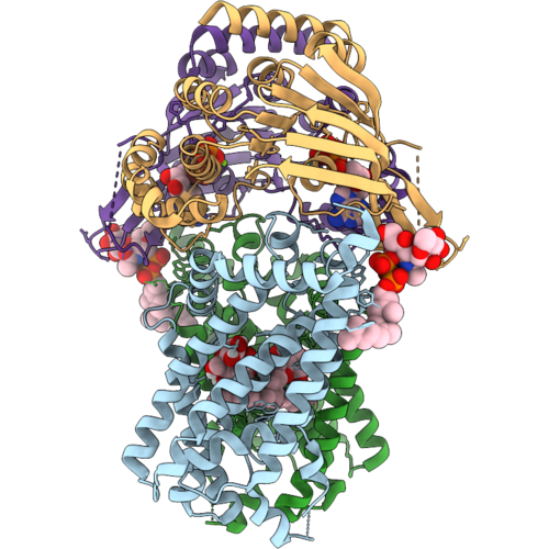
Mtb Galactan Transporter Rv3781/3783, Atp-Bound
Organism: Mycobacterium tuberculosis h37rv
Method: ELECTRON MICROSCOPY Release Date: 2026-10-07 Classification: TRANSPORT PROTEIN Ligands: MG, ATP |
Organism: Mycobacterium tuberculosis h37rv
Method: ELECTRON MICROSCOPY
Release Date: 2026-10-07
Ligands: MG, ATP
 |
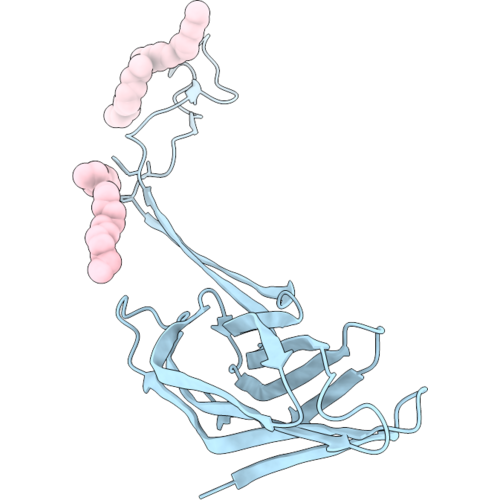
Mtb Galactan Transporter Rv3781/3783, Translocating Intermediate State
Organism: Mycobacterium tuberculosis h37rv
Method: ELECTRON MICROSCOPY Release Date: 2026-10-07 Classification: TRANSPORT PROTEIN Ligands: A1E8P, ATP, MG, LMT |
Organism: Mycobacterium tuberculosis h37rv
Method: ELECTRON MICROSCOPY
Release Date: 2026-10-07
Ligands: A1E8P, ATP, MG, LMT
 |
Mspa-M2 In A Popc Nanodisc
Organism: Mycolicibacterium smegmatis mc2 155
Method: ELECTRON MICROSCOPY Release Date: 2026-10-07 Classification: MEMBRANE PROTEIN Ligands: D12, MYS |
Organism: Mycolicibacterium smegmatis mc2 155
Method: ELECTRON MICROSCOPY
Release Date: 2026-10-07
Ligands: D12, MYS
 |
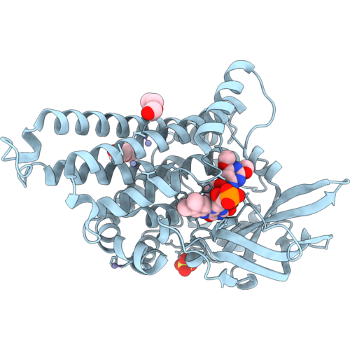
Mycobacterial Ndh-2 (Type Ii Nadh:Quinone Oxidoreductase) With Tricyclic Spirolactam Inhibitor
Organism: Mycolicibacterium smegmatis mc2 155
Method: ELECTRON MICROSCOPY Resolution:3.00 Å Release Date: 2026-09-30 Classification: MEMBRANE PROTEIN Ligands: FAD, A1DCJ |
Organism: Mycolicibacterium smegmatis mc2 155
Method: ELECTRON MICROSCOPY
Release Date: 2026-09-30
Ligands: FAD, A1DCJ
 |
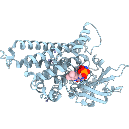
Crystal Structure Of Sulx In Complex With Fmn And Sulfadimethoxine
Organism: Microbacterium sp. cj77
Method: X-RAY DIFFRACTION Resolution:2.37 Å Release Date: 2026-09-30 Classification: OXIDOREDUCTASE Ligands: FMN, A1E1Y, ZN, PEG, SO4 |
Organism: Microbacterium sp. cj77
Method: X-RAY DIFFRACTION
Release Date: 2026-09-30
Ligands: FMN, A1E1Y, ZN, PEG, SO4
 |
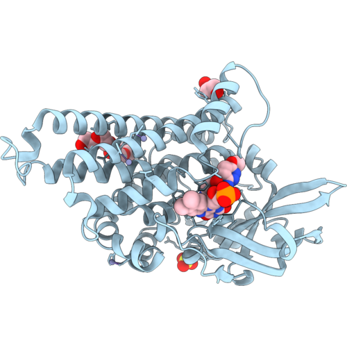
Crystal Structure Of Sulx In Complex With Fmn And Sulfanilamide
Organism: Microbacterium sp. cj77
Method: X-RAY DIFFRACTION Resolution:2.83 Å Release Date: 2026-09-30 Classification: OXIDOREDUCTASE Ligands: FMN, SAN, ZN |
Organism: Microbacterium sp. cj77
Method: X-RAY DIFFRACTION
Release Date: 2026-09-30
Ligands: FMN, SAN, ZN
 |
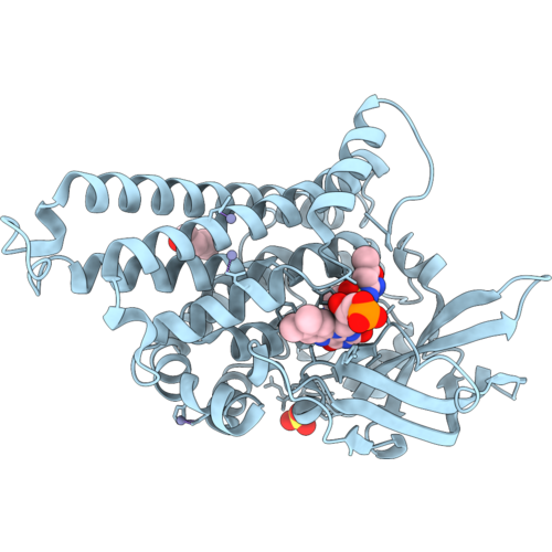
Crystal Structure Of Sulx In Complex With Fmn And Sulfamethoxypyridazine
Organism: Microbacterium sp. cj77
Method: X-RAY DIFFRACTION Resolution:2.09 Å Release Date: 2026-09-30 Classification: OXIDOREDUCTASE Ligands: FMN, PEG, SO4, PGE, ZN, A1E1X |
Organism: Microbacterium sp. cj77
Method: X-RAY DIFFRACTION
Release Date: 2026-09-30
Ligands: FMN, PEG, SO4, PGE, ZN, A1E1X
 |
Crystal Structure Of Sulx In Complex With Fmn And Sulfamethoxazole
Organism: Microbacterium sp. cj77
Method: X-RAY DIFFRACTION Resolution:2.46 Å Release Date: 2026-09-30 Classification: OXIDOREDUCTASE Ligands: PEG, FMN, SO4, ZN, 08D |
Organism: Microbacterium sp. cj77
Method: X-RAY DIFFRACTION
Release Date: 2026-09-30
Ligands: PEG, FMN, SO4, ZN, 08D