

Search Count: 53
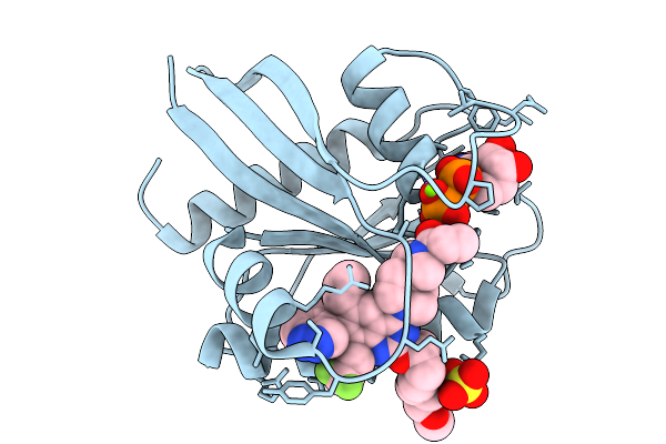 |
Krasg12C In Complex With Asp2453
Organism: Homo sapiens
Method: X-RAY DIFFRACTION Resolution:1.75 Å Release Date: 2026-03-04 Classification: HYDROLASE Ligands: GDP, MG, A1L9E, SO4 |
Organism: Homo sapiens
Method: X-RAY DIFFRACTION
Release Date: 2026-03-04
Ligands: GDP, MG, A1L9E, SO4
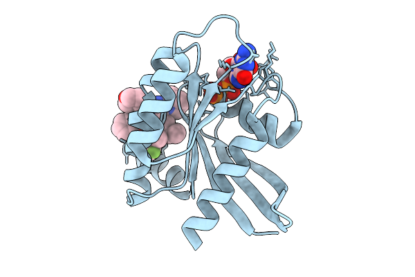 |
Crystal Structure Of Kras G12D (Gdp) In Complex With Compound 1
Organism: Homo sapiens
Method: X-RAY DIFFRACTION Resolution:1.36 Å Release Date: 2025-09-03 Classification: HYDROLASE Ligands: GDP, MG, A1L65 |
Organism: Homo sapiens
Method: X-RAY DIFFRACTION
Release Date: 2025-09-03
Ligands: GDP, MG, A1L65
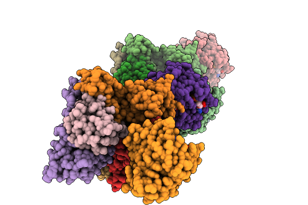 |
Crystal Structure Of Kras G12D (Gdp) In Complex With Asp3082
Organism: Homo sapiens
Method: X-RAY DIFFRACTION Resolution:3.18 Å Release Date: 2025-09-03 Classification: HYDROLASE Ligands: A1L66, GDP, MG |
Organism: Homo sapiens
Method: X-RAY DIFFRACTION
Release Date: 2025-09-03
Ligands: A1L66, GDP, MG
 |
Krasg12C In Complex With Inhibitor
Organism: Homo sapiens
Method: X-RAY DIFFRACTION Resolution:1.85 Å Release Date: 2024-01-17 Classification: HYDROLASE/INHIBITOR Ligands: GDP, MG, Y9D |
Organism: Homo sapiens
Method: X-RAY DIFFRACTION
Release Date: 2024-01-17
Ligands: GDP, MG, Y9D
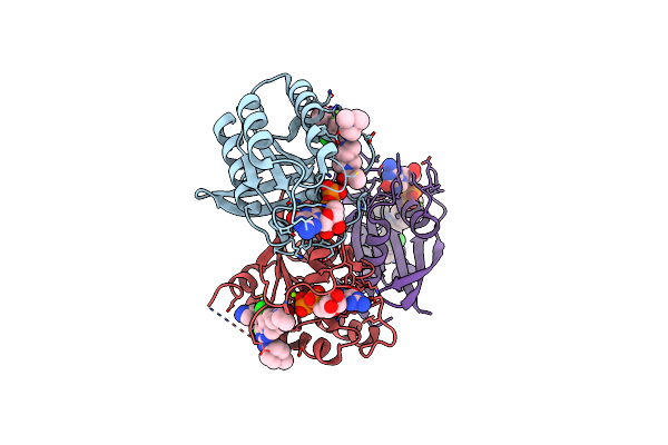 |
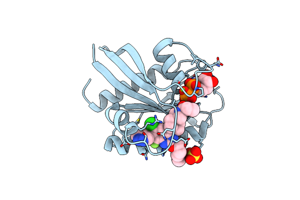
Kras G12C In Complex With Compound 5C
Organism: Homo sapiens
Method: X-RAY DIFFRACTION Resolution:1.79 Å Release Date: 2022-08-10 Classification: ONCOPROTEIN/INHIBITOR Ligands: GDP, MG, IQC |
Organism: Homo sapiens
Method: X-RAY DIFFRACTION
Release Date: 2022-08-10
Ligands: GDP, MG, IQC
 |
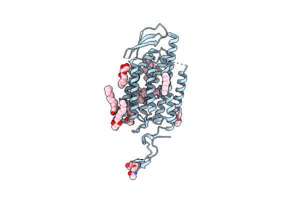
Kras G12C In Complex With Compound 7B
Organism: Homo sapiens
Method: X-RAY DIFFRACTION Resolution:1.80 Å Release Date: 2022-08-10 Classification: ONCOPROTEIN/inhibitor Ligands: GDP, MG, SO4, IQN |
Organism: Homo sapiens
Method: X-RAY DIFFRACTION
Release Date: 2022-08-10
Ligands: GDP, MG, SO4, IQN
 |
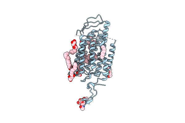
Time-Resolved Serial Femtosecond Crystallography Reveals Early Structural Changes In Channelrhodopsin: Dark State Structure
Organism: Chlamydomonas reinhardtii
Method: X-RAY DIFFRACTION Resolution:2.30 Å Release Date: 2021-04-07 Classification: MEMBRANE PROTEIN Ligands: RET, OLC |
Organism: Chlamydomonas reinhardtii
Method: X-RAY DIFFRACTION
Release Date: 2021-04-07
Ligands: RET, OLC
 |
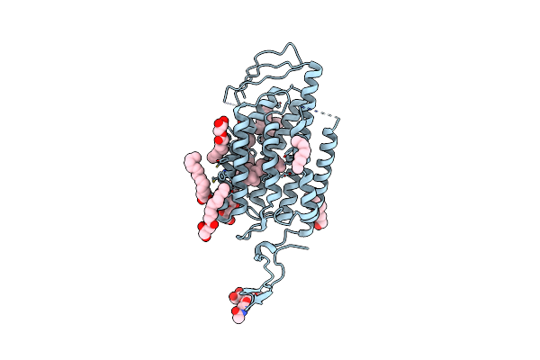
Time-Resolved Serial Femtosecond Crystallography Reveals Early Structural Changes In Channelrhodopsin: 4 Ms Structure
Organism: Chlamydomonas reinhardtii
Method: X-RAY DIFFRACTION Resolution:2.50 Å Release Date: 2021-04-07 Classification: MEMBRANE PROTEIN Ligands: RET, OLC |
Organism: Chlamydomonas reinhardtii
Method: X-RAY DIFFRACTION
Release Date: 2021-04-07
Ligands: RET, OLC
 |
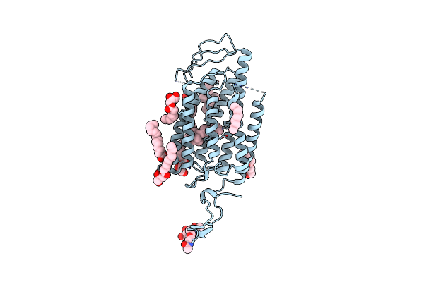
Time-Resolved Serial Femtosecond Crystallography Reveals Early Structural Changes In Channelrhodopsin: 1 Microsecond Structure
Organism: Chlamydomonas reinhardtii
Method: X-RAY DIFFRACTION Resolution:2.50 Å Release Date: 2021-04-07 Classification: MEMBRANE PROTEIN Ligands: RET, OLC |
Organism: Chlamydomonas reinhardtii
Method: X-RAY DIFFRACTION
Release Date: 2021-04-07
Ligands: RET, OLC
 |
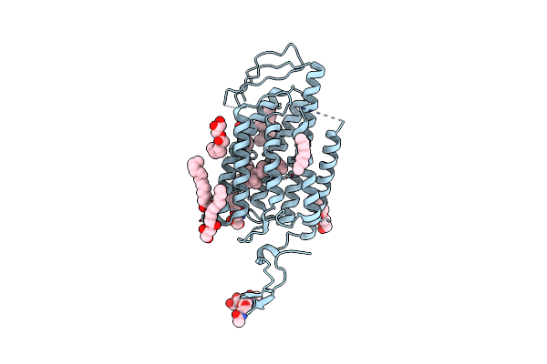
Time-Resolved Serial Femtosecond Crystallography Reveals Early Structural Changes In Channelrhodopsin: 50 Microsecond Structure
Organism: Chlamydomonas reinhardtii
Method: X-RAY DIFFRACTION Resolution:2.50 Å Release Date: 2021-04-07 Classification: MEMBRANE PROTEIN Ligands: RET, OLC |
Organism: Chlamydomonas reinhardtii
Method: X-RAY DIFFRACTION
Release Date: 2021-04-07
Ligands: RET, OLC
 |
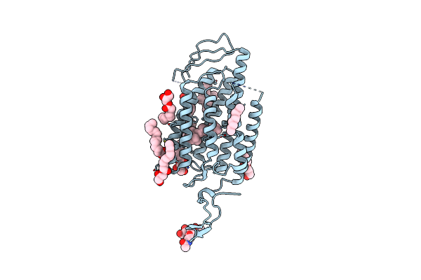
Time-Resolved Serial Femtosecond Crystallography Reveals Early Structural Changes In Channelrhodopsin: 250 Microsecond Structure
Organism: Chlamydomonas reinhardtii
Method: X-RAY DIFFRACTION Resolution:2.50 Å Release Date: 2021-04-07 Classification: MEMBRANE PROTEIN Ligands: RET, OLC |
Organism: Chlamydomonas reinhardtii
Method: X-RAY DIFFRACTION
Release Date: 2021-04-07
Ligands: RET, OLC
 |
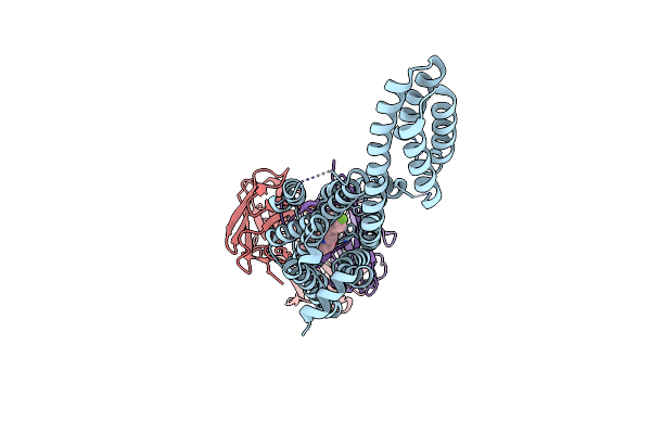
Time-Resolved Serial Femtosecond Crystallography Reveals Early Structural Changes In Channelrhodopsin: 1 Ms Structure
Organism: Chlamydomonas reinhardtii
Method: X-RAY DIFFRACTION Resolution:2.50 Å Release Date: 2021-04-07 Classification: MEMBRANE PROTEIN Ligands: RET, OLC |
Organism: Chlamydomonas reinhardtii
Method: X-RAY DIFFRACTION
Release Date: 2021-04-07
Ligands: RET, OLC
 |
Human Dopamine D2 Receptor In Complex With Spiperone
Organism: Homo sapiens, Escherichia coli, Mus musculus
Method: X-RAY DIFFRACTION Resolution:3.10 Å Release Date: 2020-12-30 Classification: MEMBRANE PROTEIN Ligands: SIP |
Organism: Homo sapiens, Escherichia coli, Mus musculus
Method: X-RAY DIFFRACTION
Release Date: 2020-12-30
Ligands: SIP
 |
Structure Of Human A2A Adenosine Receptor In Complex With Zm241385 Obtained From Sfx Experiments Under Atmospheric Pressure
Organism: Homo sapiens, Escherichia coli
Method: X-RAY DIFFRACTION Resolution:2.25 Å Release Date: 2019-10-30 Classification: MEMBRANE PROTEIN |
Organism: Homo sapiens, Escherichia coli
Method: X-RAY DIFFRACTION
Release Date: 2019-10-30
 |
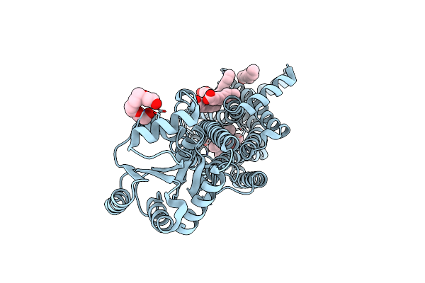
Structure Of Hen Egg-White Lysozyme Obtained From Sfx Experiments Under Atmospheric Pressure
Organism: Gallus gallus
Method: X-RAY DIFFRACTION Resolution:2.00 Å Release Date: 2019-10-30 Classification: HYDROLASE |
Organism: Gallus gallus
Method: X-RAY DIFFRACTION
Release Date: 2019-10-30
 |
Crystal Structures Of Human Orexin 2 Receptor Bound To The Selective Antagonist Empa Determined By Serial Femtosecond Crystallography At Sacla
Organism: Homo sapiens, Pyrococcus abyssi (strain ge5 / orsay)
Method: X-RAY DIFFRACTION Resolution:2.30 Å Release Date: 2017-12-13 Classification: SIGNALING PROTEIN Ligands: 7MA, OLA, 1PE |
Organism: Homo sapiens, Pyrococcus abyssi (strain ge5 / orsay)
Method: X-RAY DIFFRACTION
Release Date: 2017-12-13
Ligands: 7MA, OLA, 1PE
 |
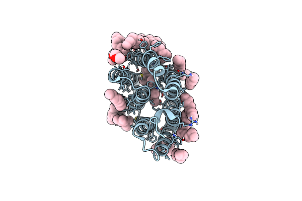
Crystal Structure Of Human Orexin 2 Receptor Bound To The Selective Antagonist Empa Determined By The Synchrotron Light Source At Spring-8.
Organism: Homo sapiens, Pyrococcus abyssi ge5
Method: X-RAY DIFFRACTION Resolution:1.96 Å Release Date: 2017-11-29 Classification: SIGNALING PROTEIN Ligands: 7MA, OLA, 1PE |
Organism: Homo sapiens, Pyrococcus abyssi ge5
Method: X-RAY DIFFRACTION
Release Date: 2017-11-29
Ligands: 7MA, OLA, 1PE
 |
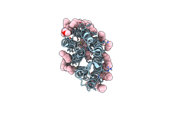
A Three Dimensional Movie Of Structural Changes In Bacteriorhodopsin: Resting State Structure
Organism: Halobacterium salinarum (strain atcc 700922 / jcm 11081 / nrc-1)
Method: X-RAY DIFFRACTION Resolution:2.00 Å Release Date: 2016-12-21 Classification: PROTON TRANSPORT Ligands: RET, L2P, TRD, D10, HP6, OCT, MYS, UND, DD9, C14 |
Organism: Halobacterium salinarum (strain atcc 700922 / jcm 11081 / nrc-1)
Method: X-RAY DIFFRACTION
Release Date: 2016-12-21
Ligands: RET, L2P, TRD, D10, HP6, OCT, MYS, UND, DD9, C14
 |
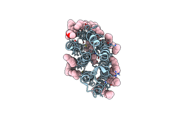
A Three Dimensional Movie Of Structural Changes In Bacteriorhodopsin: Structure Obtained 16 Ns After Photoexcitation
Organism: Halobacterium salinarum (strain atcc 700922 / jcm 11081 / nrc-1)
Method: X-RAY DIFFRACTION Resolution:2.10 Å Release Date: 2016-12-21 Classification: PROTON TRANSPORT Ligands: RET, L2P, TRD, D10, HP6, OCT, MYS, UND, DD9, C14 |
Organism: Halobacterium salinarum (strain atcc 700922 / jcm 11081 / nrc-1)
Method: X-RAY DIFFRACTION
Release Date: 2016-12-21
Ligands: RET, L2P, TRD, D10, HP6, OCT, MYS, UND, DD9, C14
 |
A Three Dimensional Movie Of Structural Changes In Bacteriorhodopsin: Structure Obtained 760 Ns After Photoexcitation
Organism: Halobacterium salinarum (strain atcc 700922 / jcm 11081 / nrc-1)
Method: X-RAY DIFFRACTION Resolution:2.10 Å Release Date: 2016-12-21 Classification: PROTON TRANSPORT Ligands: RET, L2P, TRD, D10, HP6, OCT, MYS, UND, DD9, C14 |
Organism: Halobacterium salinarum (strain atcc 700922 / jcm 11081 / nrc-1)
Method: X-RAY DIFFRACTION
Release Date: 2016-12-21
Ligands: RET, L2P, TRD, D10, HP6, OCT, MYS, UND, DD9, C14