
Search Count: 73
 |
Organism: Homo sapiens
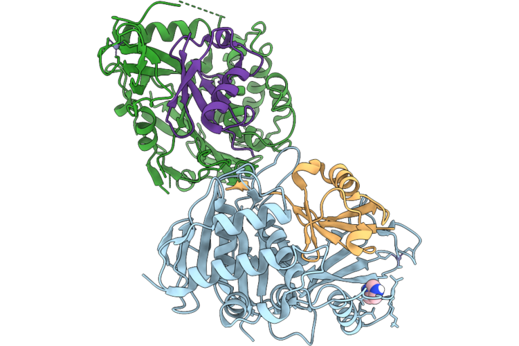
Method: X-RAY DIFFRACTION Resolution:2.30 Å Release Date: 2026-06-24 Classification: LYASE Ligands: ZN, IMD |
 |
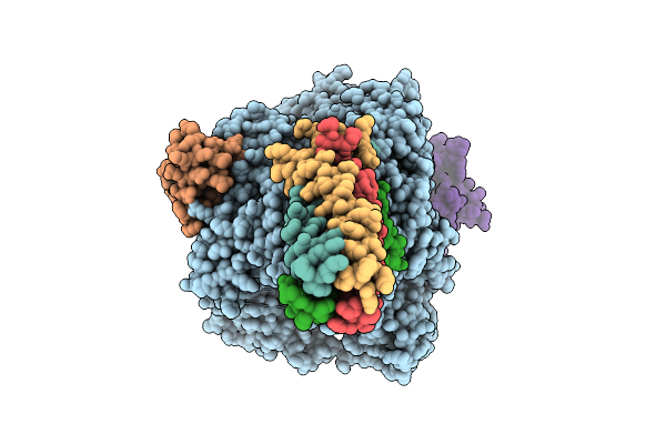
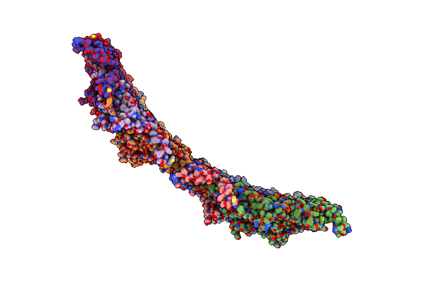
Organism: Klebsiella phage ran69
Method: ELECTRON MICROSCOPY Release Date: 2026-05-27 Classification: VIRUS |
 |
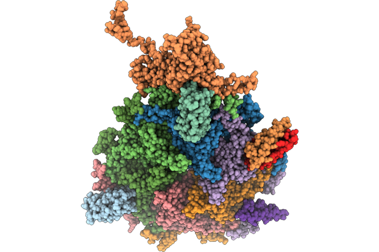
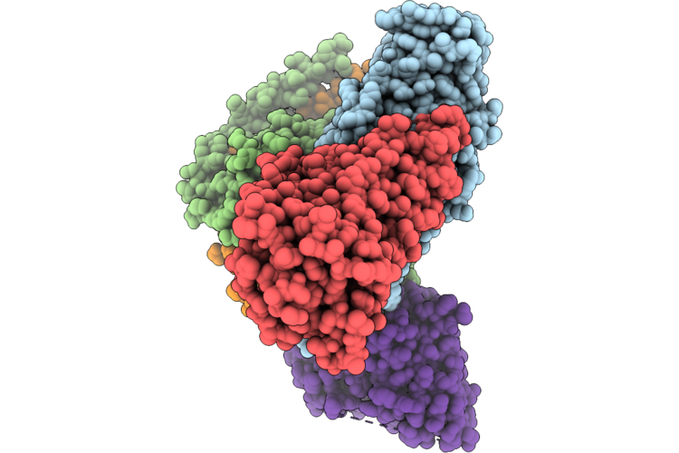
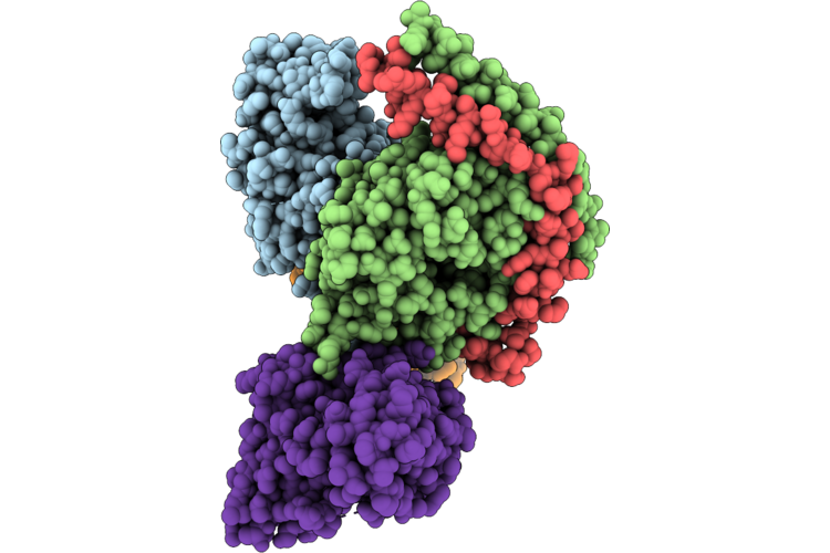
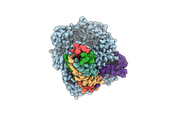
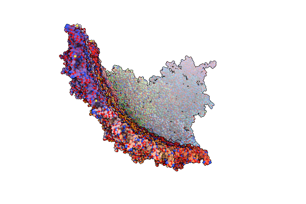
The Composite Cryo-Em Structure Of Bacteriophage Ran69 Pre-Ejectosome-Portal Complex
Organism: Klebsiella phage ran69
Method: ELECTRON MICROSCOPY Release Date: 2026-05-27 Classification: VIRUS |
 |
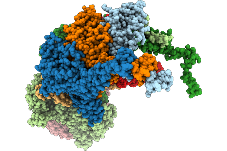
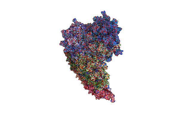
Organism: Klebsiella phage ran69
Method: ELECTRON MICROSCOPY Resolution:3.20 Å Release Date: 2026-05-27 Classification: VIRUS |
 |
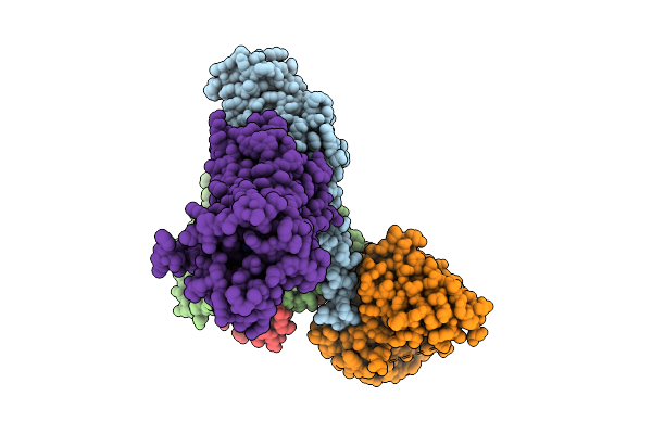
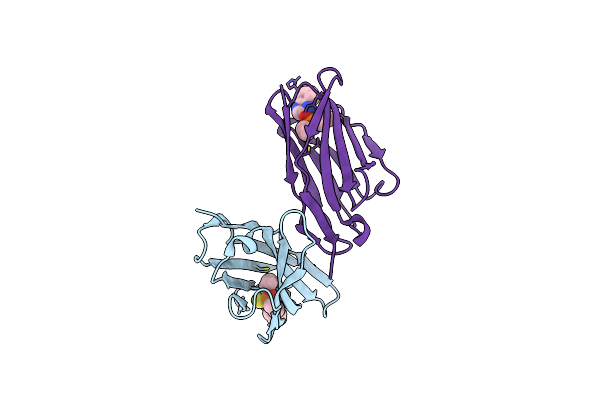
Organism: Homo sapiens, Mus musculus
Method: ELECTRON MICROSCOPY Resolution:2.42 Å Release Date: 2026-05-20 Classification: MEMBRANE PROTEIN/IMMUNE SYSTEM Ligands: A1E5S |
 |
Organism: Homo sapiens, Mus musculus
Method: ELECTRON MICROSCOPY Resolution:2.42 Å Release Date: 2026-05-20 Classification: MEMBRANE PROTEIN/IMMUNE SYSTEM Ligands: A1E5T |
 |
Solution Structures Of Brd9 Bromodomain In Complex With Histone H3 Acetyl-Lysine 18 (H3K18Ac) Peptide
Organism: Homo sapiens
Method: SOLUTION NMR Release Date: 2026-03-11 Classification: PROTEIN BINDING |
 |
Solution Structures Of Brd9 Bromodomain In Complex With Histone H3 Lactyl-Lysine 18 (H3K18La) Peptide
Organism: Homo sapiens
Method: SOLUTION NMR Release Date: 2026-03-11 Classification: PROTEIN BINDING |
 |
Organism: Homo sapiens, Mus musculus, Escherichia coli
Method: ELECTRON MICROSCOPY Resolution:3.13 Å Release Date: 2025-11-26 Classification: MEMBRANE PROTEIN Ligands: 8K3 |
 |
Organism: Mycoplasma pneumoniae (strain atcc 29342 / m129 / subtype 1)
Method: X-RAY DIFFRACTION Resolution:2.24 Å Release Date: 2025-10-29 Classification: BIOSYNTHETIC PROTEIN Ligands: GDP, MG |
 |
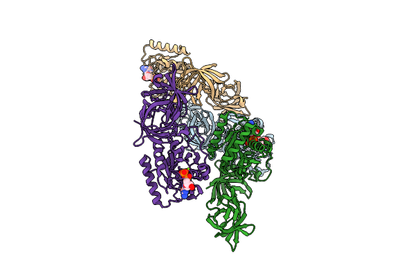
Organism: Henipavirus nipahense
Method: ELECTRON MICROSCOPY Release Date: 2025-07-30 Classification: TRANSFERASE |
 |
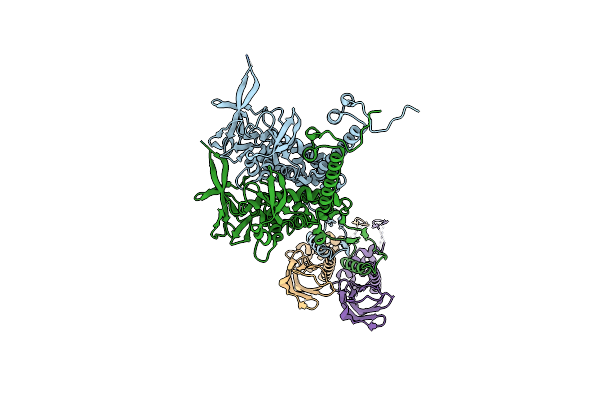
Cryo-Em Structure Of The Nipah Virus Polymerase Containing The Connecting Domain
Organism: Henipavirus nipahense
Method: ELECTRON MICROSCOPY Release Date: 2025-07-30 Classification: VIRAL PROTEIN, TRANSFERASE |
 |
Organism: Henipavirus nipahense
Method: ELECTRON MICROSCOPY Release Date: 2025-07-23 Classification: VIRAL PROTEIN, TRANSFERASE |
 |
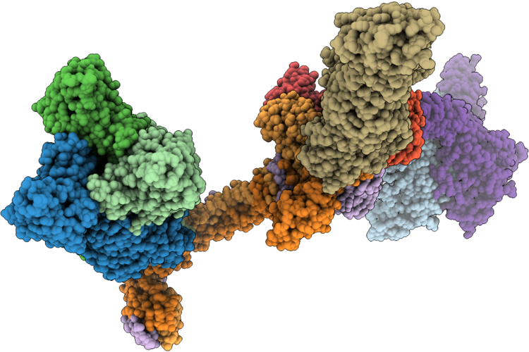
Organism: Mycolicibacterium phage mycofy1
Method: ELECTRON MICROSCOPY Release Date: 2025-04-16 Classification: VIRAL PROTEIN |
 |
Organism: Mycolicibacterium phage mycofy1
Method: ELECTRON MICROSCOPY Release Date: 2025-04-16 Classification: VIRAL PROTEIN |
 |
Organism: Mycolicibacterium phage mycofy1
Method: ELECTRON MICROSCOPY Release Date: 2025-04-16 Classification: VIRAL PROTEIN |
 |
Organism: Mycolicibacterium phage mycofy1
Method: ELECTRON MICROSCOPY Release Date: 2025-04-16 Classification: VIRAL PROTEIN |
 |
Organism: Mycolicibacterium phage mycofy1
Method: ELECTRON MICROSCOPY Release Date: 2025-04-16 Classification: VIRAL PROTEIN |
 |
Organism: Homo sapiens
Method: X-RAY DIFFRACTION Resolution:2.15 Å Release Date: 2024-07-17 Classification: STRUCTURAL GENOMICS Ligands: NO, VAL, CL, RU, PLM, GOL, DMS |
 |
Organism: Camelus bactrianus
Method: X-RAY DIFFRACTION Resolution:2.40 Å Release Date: 2023-10-25 Classification: IMMUNE SYSTEM Ligands: WYN |