

Search Count: 23
 |
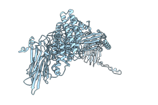
Native Structure Of Full-Length Pesticidal Protein Cry1Ca18 At Ph9, From Crystals Formed In Vivo
Organism: Bacillus thuringiensis
Method: X-RAY DIFFRACTION Resolution:1.65 Å Release Date: 2026-02-11 Classification: TOXIN Ligands: CA, ZN |
Organism: Bacillus thuringiensis
Method: X-RAY DIFFRACTION
Release Date: 2026-02-11
Ligands: CA, ZN
 |
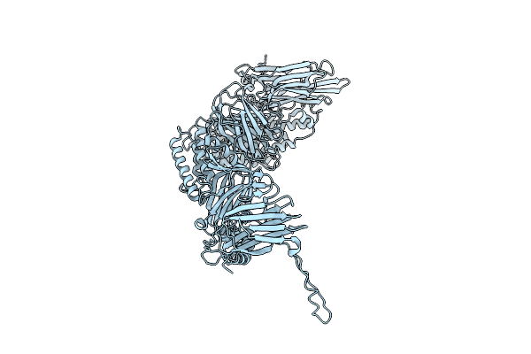
Native Structure Of The Full-Length Pesticidal Protein Cry8Ba2, From Crystals Formed In Vivo (Form 1)
Organism: Bacillus thuringiensis
Method: X-RAY DIFFRACTION Resolution:2.27 Å Release Date: 2025-12-03 Classification: TOXIN |
Organism: Bacillus thuringiensis
Method: X-RAY DIFFRACTION
Release Date: 2025-12-03
 |
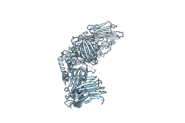
Native Structure Of The Full-Length Pesticidal Protein Cry8Ba2, From Crystals Formed In Vivo (Form 2)
Organism: Bacillus thuringiensis
Method: X-RAY DIFFRACTION Resolution:2.27 Å Release Date: 2025-12-03 Classification: TOXIN |
Organism: Bacillus thuringiensis
Method: X-RAY DIFFRACTION
Release Date: 2025-12-03
 |
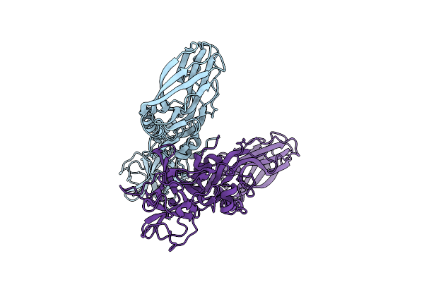
Native Structure Of The Full-Length Pesticidal Protein Cry1Ca18 At Ph7, From Crystals Formed In Vivo
Organism: Bacillus thuringiensis
Method: X-RAY DIFFRACTION Resolution:1.80 Å Release Date: 2025-11-12 Classification: TOXIN Ligands: ZN, CA |
Organism: Bacillus thuringiensis
Method: X-RAY DIFFRACTION
Release Date: 2025-11-12
Ligands: ZN, CA
 |
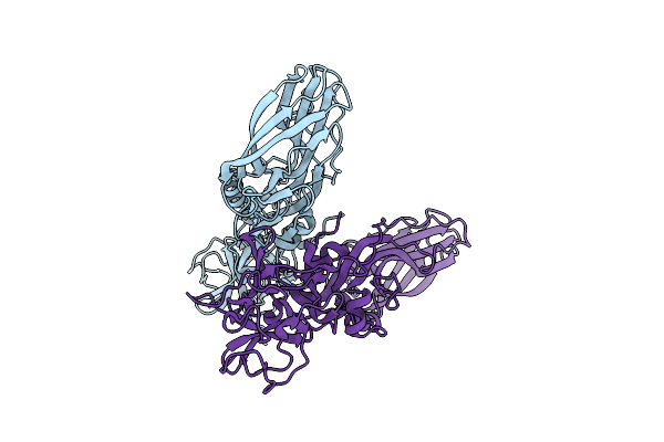
Structure Of The Lysinibacillus Sphaericus Tpp49Aa1 Pesticidal Protein At Ph 3
Organism: Lysinibacillus sphaericus
Method: X-RAY DIFFRACTION Resolution:1.78 Å Release Date: 2023-11-01 Classification: TOXIN |
Organism: Lysinibacillus sphaericus
Method: X-RAY DIFFRACTION
Release Date: 2023-11-01
 |
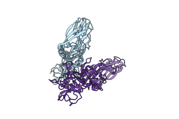
Structure Of The Lysinibacillus Sphaericus Tpp49Aa1 Pesticidal Protein At Ph 7
Organism: Lysinibacillus sphaericus
Method: X-RAY DIFFRACTION Resolution:1.62 Å Release Date: 2023-11-01 Classification: TOXIN |
Organism: Lysinibacillus sphaericus
Method: X-RAY DIFFRACTION
Release Date: 2023-11-01
 |
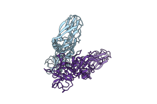
Structure Of The Lysinibacillus Sphaericus Tpp49Aa1 Pesticidal Protein At Ph 11
Organism: Lysinibacillus sphaericus
Method: X-RAY DIFFRACTION Resolution:1.75 Å Release Date: 2023-11-01 Classification: TOXIN |
Organism: Lysinibacillus sphaericus
Method: X-RAY DIFFRACTION
Release Date: 2023-11-01
 |
The Structure Of Natural Crystals Of The Lysinibacillus Sphaericus Tpp49Aa1 Pesticidal Protein Elucidated Using Serial Femtosecond Crystallography At An X-Ray Free Electron Laser
Organism: Lysinibacillus sphaericus
Method: X-RAY DIFFRACTION Resolution:2.20 Å Release Date: 2023-05-17 Classification: TOXIN |
Organism: Lysinibacillus sphaericus
Method: X-RAY DIFFRACTION
Release Date: 2023-05-17
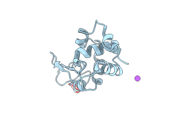 |
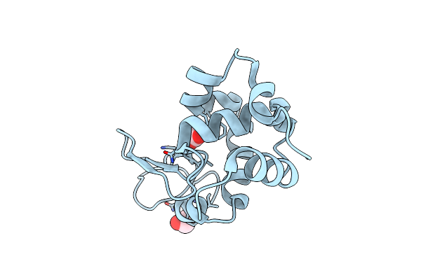
Multi-Hit Sfx Using Mhz Xfel Sources- First Hit
Organism: Gallus gallus
Method: X-RAY DIFFRACTION Resolution:3.20 Å Release Date: 2022-07-20 Classification: HYDROLASE Ligands: EDO, NA |
Organism: Gallus gallus
Method: X-RAY DIFFRACTION
Release Date: 2022-07-20
Ligands: EDO, NA
 |
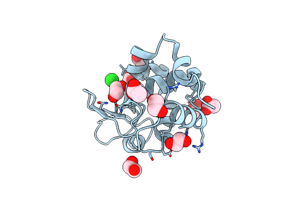
Multi-Hit Sfx Using Mhz Xfel Sources
Organism: Gallus gallus
Method: X-RAY DIFFRACTION Resolution:2.10 Å Release Date: 2021-10-13 Classification: HYDROLASE Ligands: ACT, EDO, CL |
Organism: Gallus gallus
Method: X-RAY DIFFRACTION
Release Date: 2021-10-13
Ligands: ACT, EDO, CL
 |
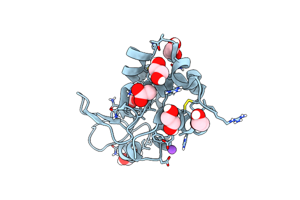
Multi-Hit Sfx Using Mhz Xfel Sources
Organism: Gallus gallus
Method: X-RAY DIFFRACTION Resolution:2.10 Å Release Date: 2021-10-13 Classification: HYDROLASE Ligands: CL, EDO, ACT |
Organism: Gallus gallus
Method: X-RAY DIFFRACTION
Release Date: 2021-10-13
Ligands: CL, EDO, ACT
 |
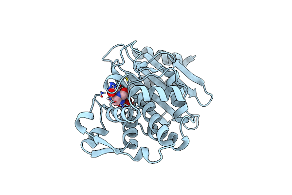
Serial Femtosecond Crystallography At Megahertz Pulse Rates
Organism: Gallus gallus
Method: X-RAY DIFFRACTION Resolution:1.76 Å Release Date: 2018-10-10 Classification: HYDROLASE Ligands: CL, NA, ACT, EDO |
Organism: Gallus gallus
Method: X-RAY DIFFRACTION
Release Date: 2018-10-10
Ligands: CL, NA, ACT, EDO
 |
Serial Femtosecond Crystallography At Megahertz Pulse Rates
Organism: Klebsiella pneumoniae
Method: X-RAY DIFFRACTION Resolution:1.69 Å Release Date: 2018-10-10 Classification: ANTIBIOTIC Ligands: NXL |
Organism: Klebsiella pneumoniae
Method: X-RAY DIFFRACTION
Release Date: 2018-10-10
Ligands: NXL
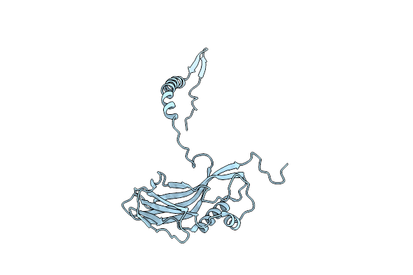 |
Sfx Structure Of Cydia Pomonella Granulovirus Using A Double Flow-Focusing Nozzle
Organism: Cydia pomonella granulosis virus (isolate mexico/1963)
Method: X-RAY DIFFRACTION Resolution:2.56 Å Release Date: 2017-03-29 Classification: VIRAL PROTEIN |
Organism: Cydia pomonella granulosis virus (isolate mexico/1963)
Method: X-RAY DIFFRACTION
Release Date: 2017-03-29
 |
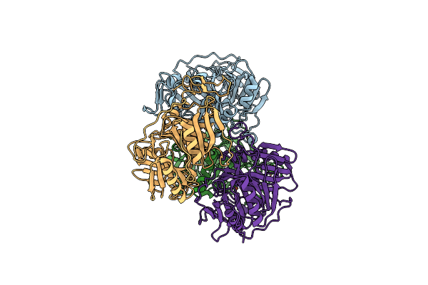
12 Subunit Rna Polymerase Ii At Room Temperature Collected Using Sfx
Organism: Saccharomyces cerevisiae (strain atcc 204508 / s288c)
Method: X-RAY DIFFRACTION Resolution:3.80 Å Release Date: 2017-03-29 Classification: TRANSFERASE Ligands: ZN, MG |
Organism: Saccharomyces cerevisiae (strain atcc 204508 / s288c)
Method: X-RAY DIFFRACTION
Release Date: 2017-03-29
Ligands: ZN, MG
 |
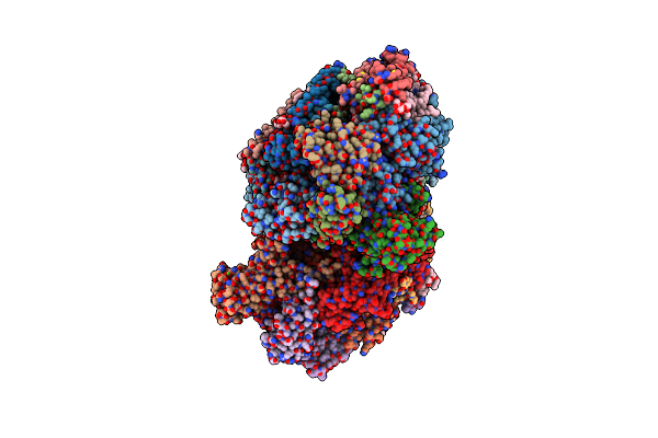
Room Temperature Structure Of An Extradiol Ring-Cleaving Dioxygenase From B.Fuscum Determined Using Serial Femtosecond Crystallography
Organism: Brevibacterium fuscum
Method: X-RAY DIFFRACTION Resolution:2.38 Å Release Date: 2017-03-22 Classification: OXIDOREDUCTASE Ligands: FE2, CL, CA |
Organism: Brevibacterium fuscum
Method: X-RAY DIFFRACTION
Release Date: 2017-03-22
Ligands: FE2, CL, CA
 |
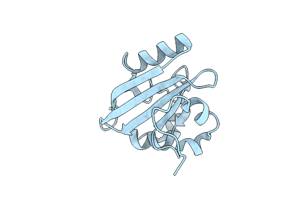
Macromolecular Diffractive Imaging Using Imperfect Crystals
Organism: Thermosynechococcus elongatus (strain bp-1)
Method: X-RAY DIFFRACTION Resolution:3.50 Å Release Date: 2017-02-08 Classification: PHOTOSYNTHESIS Ligands: OEX, FE2, CL, BCT, CLA, PHO, BCR, PL9, SQD, LMG, LHG, CA, DGD, HEM, MG |
 |
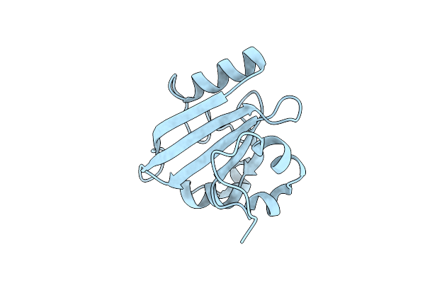
Femtosecond Structural Dynamics Drives The Trans/Cis Isomerization In Photoactive Yellow Protein: 200 Ns Time Delay Photo-Activated (Light) Structure
Organism: Halorhodospira halophila
Method: X-RAY DIFFRACTION Resolution:1.60 Å Release Date: 2016-05-25 Classification: SIGNALING PROTEIN |
Organism: Halorhodospira halophila
Method: X-RAY DIFFRACTION
Release Date: 2016-05-25
 |
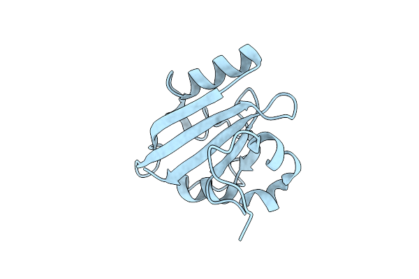
Femtosecond Structural Dynamics Drives The Trans/Cis Isomerization In Photoactive Yellow Protein: 100 Fs To 400 Fs Structure
Organism: Halorhodospira halophila
Method: X-RAY DIFFRACTION Resolution:1.60 Å Release Date: 2016-05-25 Classification: SIGNALING PROTEIN |
Organism: Halorhodospira halophila
Method: X-RAY DIFFRACTION
Release Date: 2016-05-25
 |
Femtosecond Structural Dynamics Drives The Trans/Cis Isomerization In Photoactive Yellow Protein: 800 Fs To 1200 Fs Structure
Organism: Halorhodospira halophila
Method: X-RAY DIFFRACTION Resolution:1.60 Å Release Date: 2016-05-25 Classification: SIGNALING PROTEIN |
Organism: Halorhodospira halophila
Method: X-RAY DIFFRACTION
Release Date: 2016-05-25