

Search Count: 28
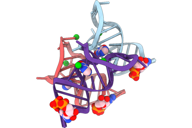 |
Crystal Structure Of Xanthine/Guanine Rna Aptamer With Xanthine
Organism: Synthetic construct
Method: X-RAY DIFFRACTION Resolution:2.51 Å Release Date: 2026-09-23 Classification: RNA Ligands: GTP, XAN, BA |
Organism: Synthetic construct
Method: X-RAY DIFFRACTION
Release Date: 2026-09-23
Ligands: GTP, XAN, BA
 |
Crystal Structure Of Xanthine/Guanine Rna Aptamer (P1 12Bp) With Xanthine
Organism: Synthetic construct
Method: X-RAY DIFFRACTION Resolution:2.19 Å Release Date: 2026-09-16 Classification: RNA Ligands: GTP, XAN, NA, GDP |
Organism: Synthetic construct
Method: X-RAY DIFFRACTION
Release Date: 2026-09-16
Ligands: GTP, XAN, NA, GDP
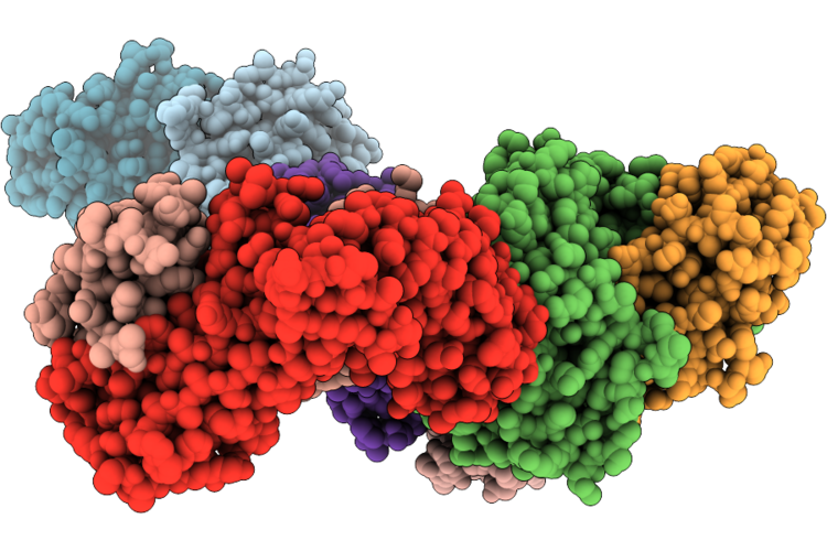 |
Crystal Structure Of Mait A-F7 Tcr-Mr1-Xanthine
Organism: Homo sapiens
Method: X-RAY DIFFRACTION Resolution:2.60 Å Release Date: 2026-08-26 Classification: IMMUNE SYSTEM Ligands: XAN, GOL, CL, CA |
Organism: Homo sapiens
Method: X-RAY DIFFRACTION
Release Date: 2026-08-26
Ligands: XAN, GOL, CL, CA
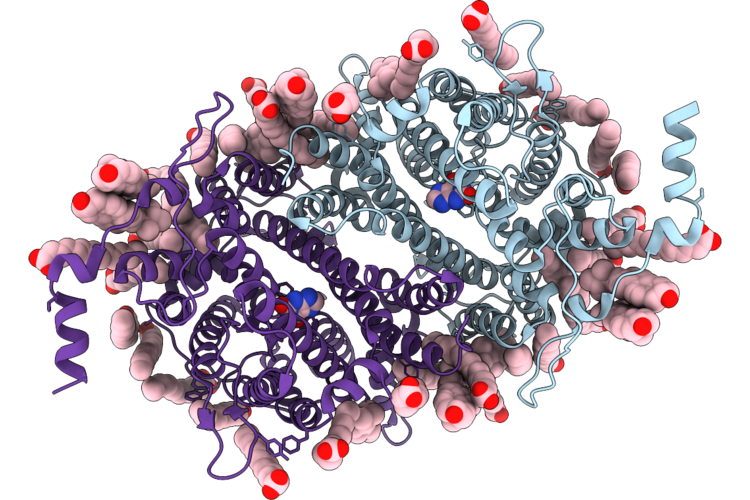 |
Uapa (Q408E) In Msp1E3D1 Nanodiscs, Xanthine-Bound State
Organism: Aspergillus nidulans fgsc a4
Method: ELECTRON MICROSCOPY Resolution:3.48 Å Release Date: 2026-07-01 Classification: MEMBRANE PROTEIN Ligands: MYR, ERG, XAN |
Organism: Aspergillus nidulans fgsc a4
Method: ELECTRON MICROSCOPY
Release Date: 2026-07-01
Ligands: MYR, ERG, XAN
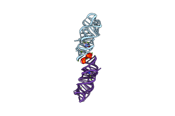 |
Crystal Structure Of P. Beijingensis Xanthine-Ii Riboswitch In Complex With Xanthine
Organism: Paenibacillus beijingensis
Method: X-RAY DIFFRACTION Resolution:2.06 Å Release Date: 2025-06-04 Classification: RNA Ligands: GTP, MG, XAN |
Organism: Paenibacillus beijingensis
Method: X-RAY DIFFRACTION
Release Date: 2025-06-04
Ligands: GTP, MG, XAN
 |
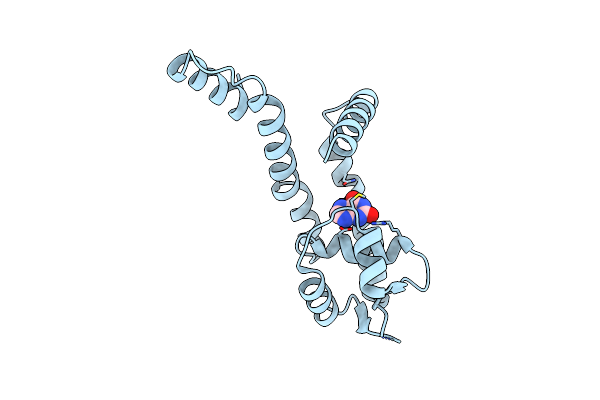
Crystal Structure Of Xanthine-Ii Ml2/3 In Complex With Xanthine
Organism: Paenibacillus beijingensis
Method: X-RAY DIFFRACTION Resolution:2.48 Å Release Date: 2025-06-04 Classification: RNA Ligands: GTP, MG, XAN |
Organism: Paenibacillus beijingensis
Method: X-RAY DIFFRACTION
Release Date: 2025-06-04
Ligands: GTP, MG, XAN
 |
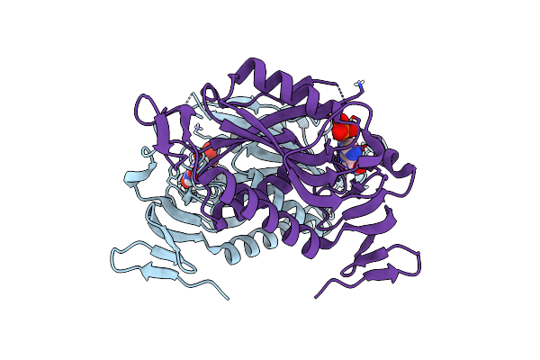
Crystal Structure Of Burkholderia Thailandensis Mftr In Complex With Xanthine
Organism: Burkholderia thailandensis
Method: X-RAY DIFFRACTION Resolution:1.80 Å Release Date: 2024-11-13 Classification: TRANSCRIPTION Ligands: XAN |
Organism: Burkholderia thailandensis
Method: X-RAY DIFFRACTION
Release Date: 2024-11-13
Ligands: XAN
 |
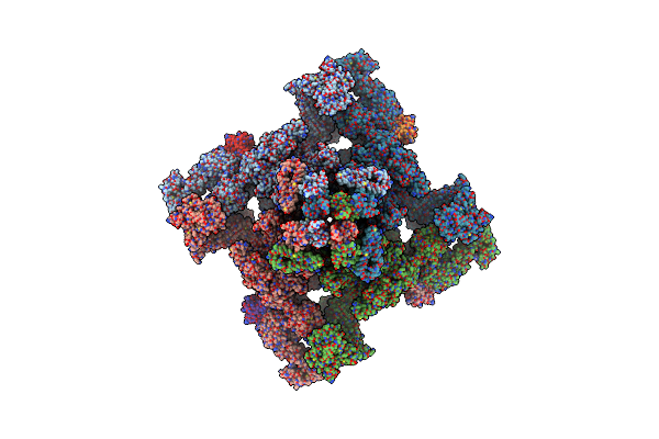
Crystal Structure Of The Trypanosoma Cruzi Hypoxanthine-Guanine-Xanthine Phosphoribosyltransferase (Hgxprt), Isoform D, Bound To The Dead-End Complex Xanthine And Pyrophosphate
Organism: Trypanosoma cruzi (strain cl brener)
Method: X-RAY DIFFRACTION Resolution:1.54 Å Release Date: 2023-07-19 Classification: TRANSFERASE Ligands: XAN, PO4 |
Organism: Trypanosoma cruzi (strain cl brener)
Method: X-RAY DIFFRACTION
Release Date: 2023-07-19
Ligands: XAN, PO4
 |
Structure Of Pka Phosphorylated Human Ryr2 In The Open State
Organism: Homo sapiens
Method: ELECTRON MICROSCOPY Release Date: 2022-08-03 Classification: MEMBRANE PROTEIN Ligands: ZN, ATP, CA, XAN |
Organism: Homo sapiens
Method: ELECTRON MICROSCOPY
Release Date: 2022-08-03
Ligands: ZN, ATP, CA, XAN
 |
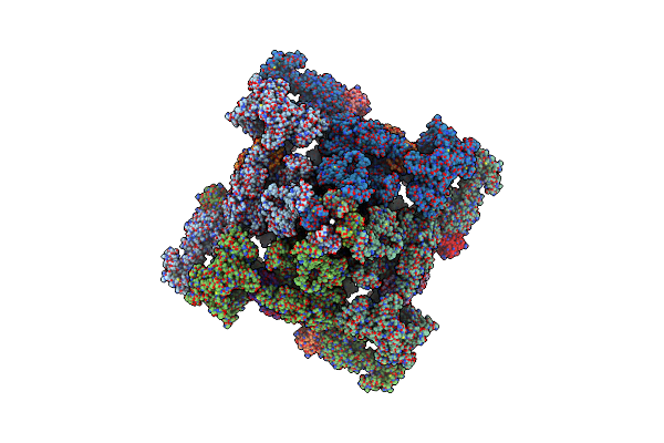
Structure Of Pka Phosphorylated Human Ryr2-R2474S In The Open State
Organism: Homo sapiens
Method: ELECTRON MICROSCOPY Release Date: 2022-08-03 Classification: MEMBRANE PROTEIN Ligands: ZN, ATP, CA, XAN |
Organism: Homo sapiens
Method: ELECTRON MICROSCOPY
Release Date: 2022-08-03
Ligands: ZN, ATP, CA, XAN
 |
Structure Of Pka Phosphorylated Human Ryr2-R2474S In The Open State In The Presence Of Calmodulin
Organism: Homo sapiens
Method: ELECTRON MICROSCOPY Release Date: 2022-08-03 Classification: MEMBRANE PROTEIN Ligands: ZN, ATP, CA, XAN |
Organism: Homo sapiens
Method: ELECTRON MICROSCOPY
Release Date: 2022-08-03
Ligands: ZN, ATP, CA, XAN
 |
Structure Of Dephosphorylated Human Ryr2 In The Open State
Organism: Homo sapiens
Method: ELECTRON MICROSCOPY Release Date: 2022-08-03 Classification: MEMBRANE PROTEIN Ligands: ZN, ATP, CA, XAN |
Organism: Homo sapiens
Method: ELECTRON MICROSCOPY
Release Date: 2022-08-03
Ligands: ZN, ATP, CA, XAN
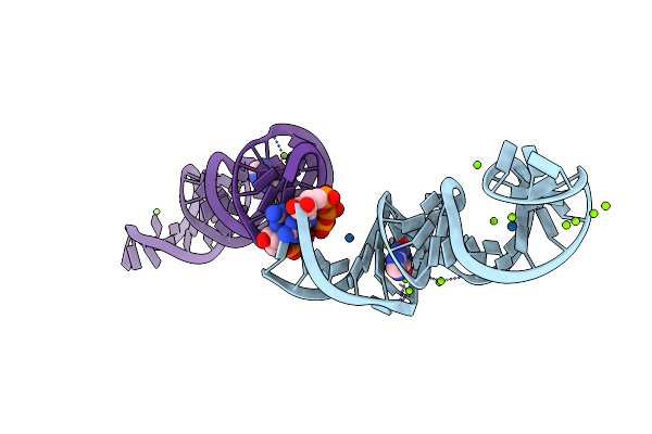 |
Crystal Structure Of Xanthine Riboswitch With Xanthine, Iridium Hexammine Soak
Organism: Ideonella sp. b508-1
Method: X-RAY DIFFRACTION Resolution:2.79 Å Release Date: 2021-06-30 Classification: RNA Ligands: GTP, IR, XAN, MG |
Organism: Ideonella sp. b508-1
Method: X-RAY DIFFRACTION
Release Date: 2021-06-30
Ligands: GTP, IR, XAN, MG
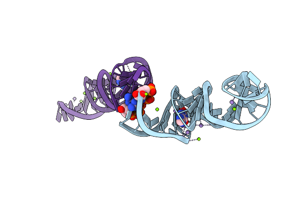 |
Crystal Structure Of Xanthine Riboswitch With Xanthine, Manganese Saok
Organism: Ideonella sp. b508-1
Method: X-RAY DIFFRACTION Resolution:2.60 Å Release Date: 2021-06-30 Classification: RNA Ligands: GTP, XAN, MN, MG |
Organism: Ideonella sp. b508-1
Method: X-RAY DIFFRACTION
Release Date: 2021-06-30
Ligands: GTP, XAN, MN, MG
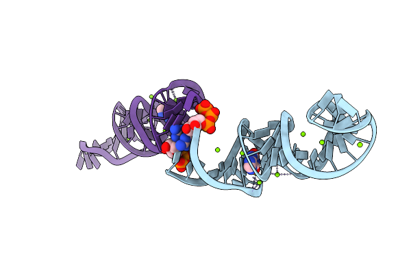 |
Crystal Structure Of Xanthine Riboswitch With Xanthine
Organism: Ideonella sp. b508-1
Method: X-RAY DIFFRACTION Resolution:2.66 Å Release Date: 2021-06-30 Classification: RNA Ligands: GTP, XAN, MG |
Organism: Ideonella sp. b508-1
Method: X-RAY DIFFRACTION
Release Date: 2021-06-30
Ligands: GTP, XAN, MG
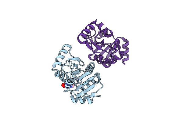 |
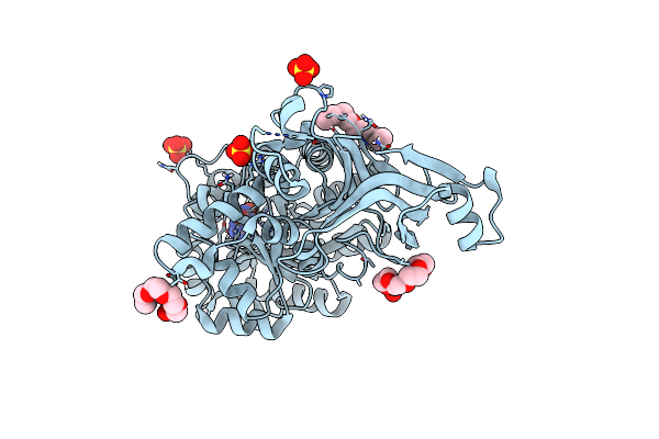
Pandda Analysis Group Deposition -- Crystal Structure Of Sars-Cov-2 Nsp3 Macrodomain In Complex With Zinc000013517187
Organism: Severe acute respiratory syndrome coronavirus 2
Method: X-RAY DIFFRACTION Resolution:1.00 Å Release Date: 2020-12-16 Classification: VIRAL PROTEIN, HYDROLASE Ligands: XAN |
Organism: Severe acute respiratory syndrome coronavirus 2
Method: X-RAY DIFFRACTION
Release Date: 2020-12-16
Ligands: XAN
 |
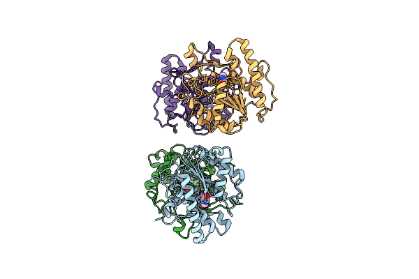
Yeast Guanine Deaminase
Organism: Saccharomyces cerevisiae (strain atcc 204508 / s288c)
Method: X-RAY DIFFRACTION Resolution:2.21 Å Release Date: 2019-07-24 Classification: HYDROLASE Ligands: XAN, ZN, 1PE, SO4 |
Organism: Saccharomyces cerevisiae (strain atcc 204508 / s288c)
Method: X-RAY DIFFRACTION
Release Date: 2019-07-24
Ligands: XAN, ZN, 1PE, SO4
 |
Crystal Structure Of Uracil Dna Glycosylase -Xanthine Complex From Bradyrhizobium Diazoefficiens
Organism: Bradyrhizobium diazoefficiens usda 110
Method: X-RAY DIFFRACTION Resolution:2.80 Å Release Date: 2017-05-03 Classification: HYDROLASE Ligands: XAN |
Organism: Bradyrhizobium diazoefficiens usda 110
Method: X-RAY DIFFRACTION
Release Date: 2017-05-03
Ligands: XAN
 |
Crystal Structure Of Geobacter Metallireducens Smug1 With Xanthine
Organism: Geobacter metallireducens (strain gs-15 / atcc 53774 / dsm 7210)
Method: X-RAY DIFFRACTION Resolution:1.50 Å Release Date: 2016-04-27 Classification: HYDROLASE Ligands: XAN, GOL, BME, IPA, LEA |
Organism: Geobacter metallireducens (strain gs-15 / atcc 53774 / dsm 7210)
Method: X-RAY DIFFRACTION
Release Date: 2016-04-27
Ligands: XAN, GOL, BME, IPA, LEA
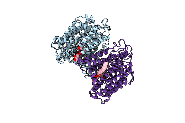 |
The Structure Of The Eukaryotic Purine/H+ Symporter, Uapa, In Complex With Xanthine
Organism: Aspergillus nidulans fgsc a4
Method: X-RAY DIFFRACTION Resolution:3.70 Å Release Date: 2016-04-27 Classification: TRANSPORT PROTEIN Ligands: XAN, LMT |
Organism: Aspergillus nidulans fgsc a4
Method: X-RAY DIFFRACTION
Release Date: 2016-04-27
Ligands: XAN, LMT