

Search Count: 41
 |
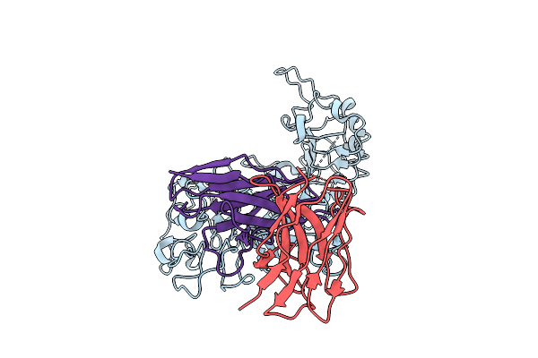
Structure Of Human Her2 In Complex With Eps232 Fab
Organism: Homo sapiens
Method: ELECTRON MICROSCOPY Resolution:3.50 Å Release Date: 2026-03-11 Classification: ANTITUMOR PROTEIN |
Organism: Homo sapiens
Method: ELECTRON MICROSCOPY
Release Date: 2026-03-11
 |
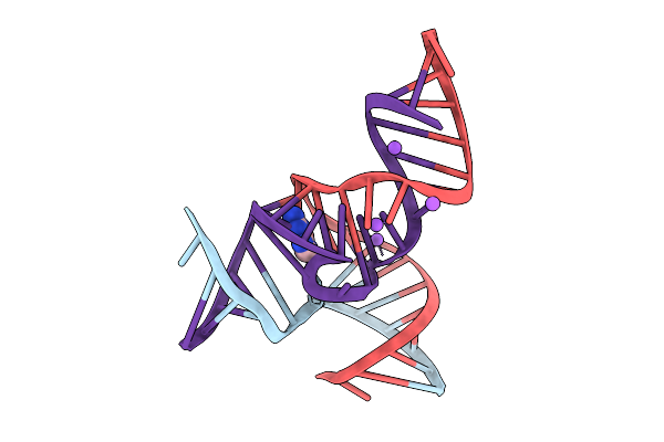
Structure Of Human Her2 In Complex With Eps226 Fab
Organism: Homo sapiens
Method: ELECTRON MICROSCOPY Resolution:3.00 Å Release Date: 2026-03-11 Classification: ANTITUMOR PROTEIN |
Organism: Homo sapiens
Method: ELECTRON MICROSCOPY
Release Date: 2026-03-11
 |
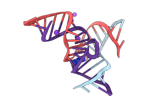
Crystal Structure Of The Methyltransferase-Ribozyme 1 Bound To Dna Substrate (With 1-Methyl-Adenosine)
Organism: Synthetic construct
Method: X-RAY DIFFRACTION Resolution:2.65 Å Release Date: 2025-09-17 Classification: DNA-RNA HYBRID |
Organism: Synthetic construct
Method: X-RAY DIFFRACTION
Release Date: 2025-09-17
 |
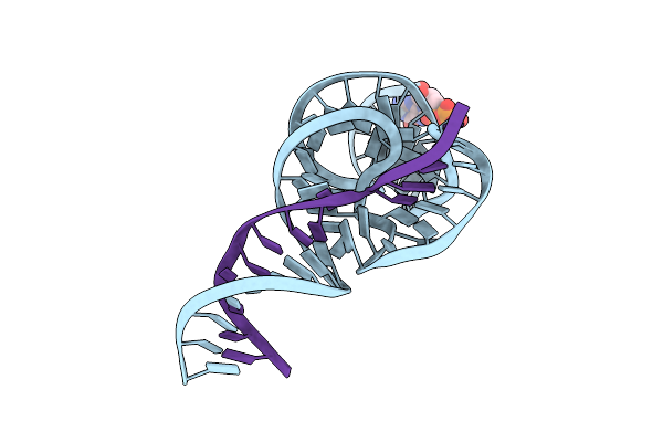
Crystal Structure Of The Methyltransferase-Ribozyme 1 Bound To Dna Substrate (1-Benzyl-Adenosine Derivative)
Organism: Synthetic construct
Method: X-RAY DIFFRACTION Resolution:2.82 Å Release Date: 2025-09-17 Classification: DNA-RNA HYBRID |
Organism: Synthetic construct
Method: X-RAY DIFFRACTION
Release Date: 2025-09-17
 |
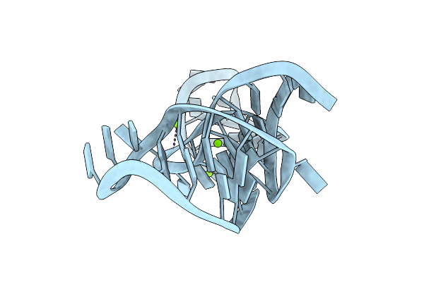
Crystal Structure Of A Hammerhead Ribozyme With Pseudoknot
Organism: Synthetic construct
Method: X-RAY DIFFRACTION Resolution:2.89 Å Release Date: 2024-08-21 Classification: RNA Ligands: GTP |
Organism: Synthetic construct
Method: X-RAY DIFFRACTION
Release Date: 2024-08-21
Ligands: GTP
 |
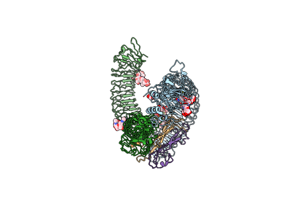
Crystal Structure Of Duf-3268 K-Junction
Organism: Bacterium
Method: X-RAY DIFFRACTION Resolution:1.94 Å Release Date: 2024-03-27 Classification: RNA Ligands: MG |
Organism: Bacterium
Method: X-RAY DIFFRACTION
Release Date: 2024-03-27
Ligands: MG
 |
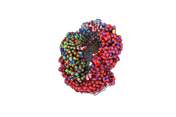
Cryo-Em Of Aedes Aegypti Toll5A Dimer Bound To Spz1C
Organism: Aedes aegypti
Method: ELECTRON MICROSCOPY Release Date: 2022-07-13 Classification: IMMUNE SYSTEM Ligands: NAG |
Organism: Aedes aegypti
Method: ELECTRON MICROSCOPY
Release Date: 2022-07-13
Ligands: NAG
 |
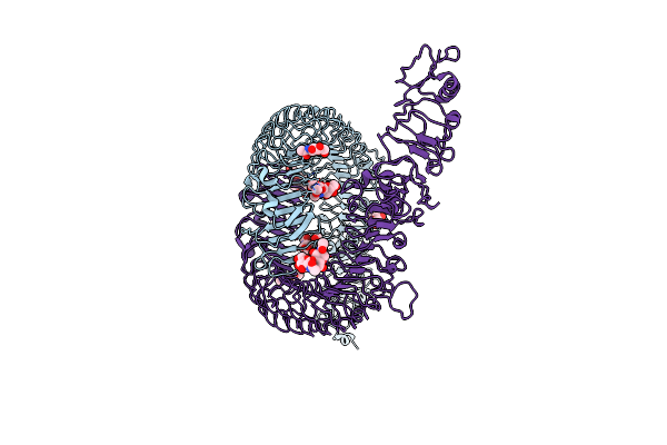
Cryo-Em Of Aedes Aegypti Toll5A Trimer Bound To Spz1C
Organism: Aedes aegypti
Method: ELECTRON MICROSCOPY Release Date: 2022-07-13 Classification: IMMUNE SYSTEM Ligands: NAG |
Organism: Aedes aegypti
Method: ELECTRON MICROSCOPY
Release Date: 2022-07-13
Ligands: NAG
 |
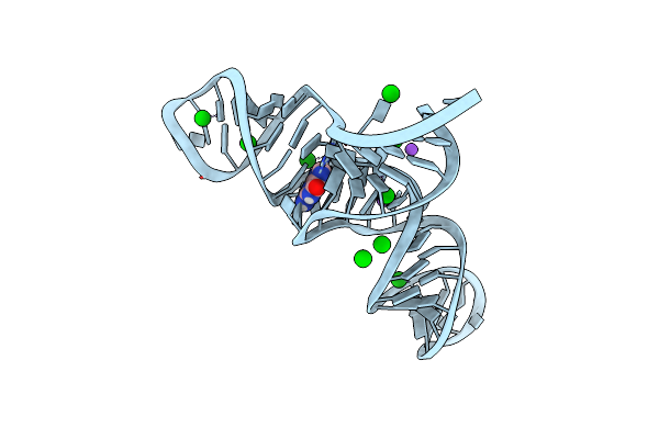
Cryo-Em Of Aedes Aegypti Toll5A
Organism: Aedes aegypti
Method: ELECTRON MICROSCOPY Release Date: 2022-07-13 Classification: IMMUNE SYSTEM Ligands: NAG |
Organism: Aedes aegypti
Method: ELECTRON MICROSCOPY
Release Date: 2022-07-13
Ligands: NAG
 |
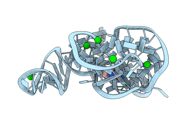
Crystal Structure Of A Methyl Transferase Ribozyme
Organism: Homo sapiens
Method: X-RAY DIFFRACTION Resolution:2.30 Å Release Date: 2022-03-23 Classification: RNA Ligands: GUN, BA, NA |
Organism: Homo sapiens
Method: X-RAY DIFFRACTION
Release Date: 2022-03-23
Ligands: GUN, BA, NA
 |
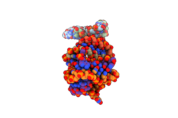
Crystal Structure Of Sam-I Riboswitch With The Actinomyces-1 K-Turn
Organism: Caldanaerobacter subterraneus subsp. tengcongensis
Method: X-RAY DIFFRACTION Resolution:2.85 Å Release Date: 2021-06-02 Classification: RNA Ligands: SAM, BA, NA |
Organism: Caldanaerobacter subterraneus subsp. tengcongensis
Method: X-RAY DIFFRACTION
Release Date: 2021-06-02
Ligands: SAM, BA, NA
 |
Crystal Structure Of The Ragath-18 K-Turn
Organism: Clostridium perfringens sm101
Method: X-RAY DIFFRACTION Resolution:2.50 Å Release Date: 2021-06-02 Classification: RNA |
Organism: Clostridium perfringens sm101
Method: X-RAY DIFFRACTION
Release Date: 2021-06-02
 |
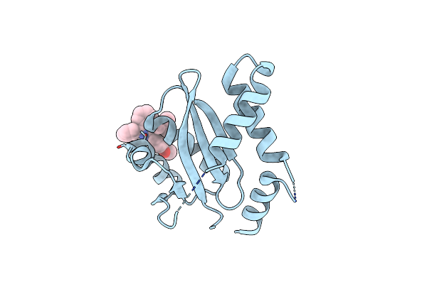
The Structure Of Pistol Ribozyme Bound To Magnesium
Organism: Alistipes putredinis
Method: X-RAY DIFFRACTION Resolution:3.10 Å Release Date: 2019-07-03 Classification: RNA Ligands: MG |
Organism: Alistipes putredinis
Method: X-RAY DIFFRACTION
Release Date: 2019-07-03
Ligands: MG
 |
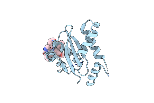
Hiv-1 Integrase Catalytic Core Domain Complexed With Allosteric Inhibitor (2S)-Tert-Butoxy[3-(3,4-Dihydro-2H-1-Benzopyran-6-Yl)-1-Phenylisoquinolin-4-Yl]Acetic Acid
Organism: Human immunodeficiency virus 1
Method: X-RAY DIFFRACTION Resolution:2.20 Å Release Date: 2019-03-06 Classification: TRANSFERASE/TRANSFERASE INHIBITOR Ligands: J3M |
Organism: Human immunodeficiency virus 1
Method: X-RAY DIFFRACTION
Release Date: 2019-03-06
Ligands: J3M
 |
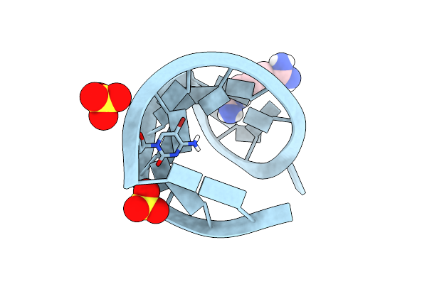
Hiv-1 Integrase Catalytic Core Domain Complexed With Allosteric Inhibitor (2S)-[1-(1-Benzyl-1H-Pyrazol-4-Yl)-3-(3,4-Dihydro-2H-1-Benzopyran-6-Yl)Isoquinolin-4-Yl](Tert-Butoxy)Acetic Acid
Organism: Human immunodeficiency virus 1
Method: X-RAY DIFFRACTION Resolution:2.49 Å Release Date: 2019-03-06 Classification: TRANSFERASE/TRANSFERASE INHIBITOR Ligands: J3P |
Organism: Human immunodeficiency virus 1
Method: X-RAY DIFFRACTION
Release Date: 2019-03-06
Ligands: J3P
 |
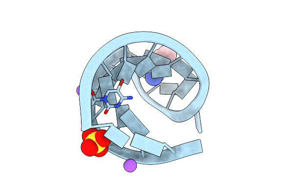
The Structure Of The G. Violaceus Guanidine Ii Riboswitch P2 Stem-Loop With Arcaine
Organism: Gloeobacter violaceus
Method: X-RAY DIFFRACTION Resolution:1.66 Å Release Date: 2019-02-20 Classification: RNA Ligands: SO4, FXQ |
Organism: Gloeobacter violaceus
Method: X-RAY DIFFRACTION
Release Date: 2019-02-20
Ligands: SO4, FXQ
 |
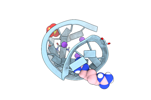
The Structure Of The G. Violaceus Guanidine Ii Riboswitch P2 Stem-Loop With Ethylguanidine
Organism: Gloeobacter violaceus
Method: X-RAY DIFFRACTION Resolution:1.54 Å Release Date: 2019-02-20 Classification: RNA Ligands: EGD, SO4, NA |
Organism: Gloeobacter violaceus
Method: X-RAY DIFFRACTION
Release Date: 2019-02-20
Ligands: EGD, SO4, NA
 |
The Structure Of The G. Violaceus Guanidine Ii Riboswitch P2 Stem-Loop With Audouine
Organism: Gloeobacter violaceus
Method: X-RAY DIFFRACTION Resolution:1.41 Å Release Date: 2019-02-20 Classification: RNA Ligands: SO4, NA, FXT |
Organism: Gloeobacter violaceus
Method: X-RAY DIFFRACTION
Release Date: 2019-02-20
Ligands: SO4, NA, FXT
 |
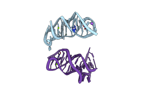
The Structure Of The Thermobifida Fusca Guanidine Iii Riboswitch With Guanidine.
Organism: Thermobifida fusca
Method: X-RAY DIFFRACTION Resolution:1.91 Å Release Date: 2017-10-18 Classification: RNA Ligands: GAI, K, NA, MG |
Organism: Thermobifida fusca
Method: X-RAY DIFFRACTION
Release Date: 2017-10-18
Ligands: GAI, K, NA, MG
 |
The Structure Of The Thermobifida Fusca Guanidine Iii Riboswitch With Aminoguanidine
Organism: Thermobifida fusca
Method: X-RAY DIFFRACTION Resolution:2.04 Å Release Date: 2017-10-18 Classification: RNA Ligands: AGU, MG, NA |
Organism: Thermobifida fusca
Method: X-RAY DIFFRACTION
Release Date: 2017-10-18
Ligands: AGU, MG, NA