

Search Count: 51
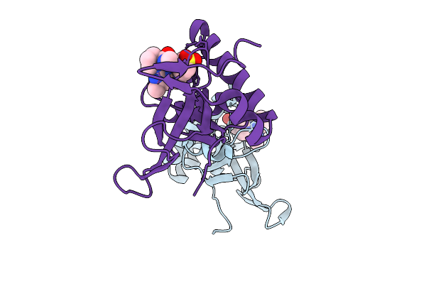 |
Crystal Structure Of The Pwwp1 Domain Of Nsd2 Bound By Compound 11.
Organism: Homo sapiens
Method: X-RAY DIFFRACTION Resolution:1.62 Å Release Date: 2025-09-03 Classification: TRANSFERASE Ligands: A1IFS, A1IEG |
Organism: Homo sapiens
Method: X-RAY DIFFRACTION
Release Date: 2025-09-03
Ligands: A1IFS, A1IEG
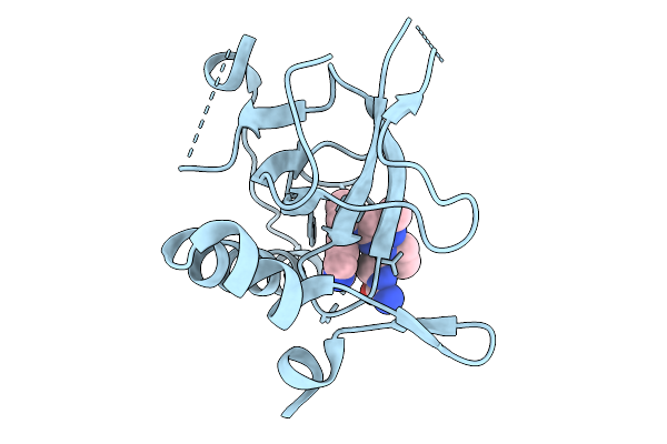 |
Crystal Structure Of The Pwwp1 Domain Of Nsd2 Bound By Compound 7.
Organism: Homo sapiens
Method: X-RAY DIFFRACTION Resolution:1.96 Å Release Date: 2025-09-03 Classification: TRANSFERASE Ligands: A1IEF |
Organism: Homo sapiens
Method: X-RAY DIFFRACTION
Release Date: 2025-09-03
Ligands: A1IEF
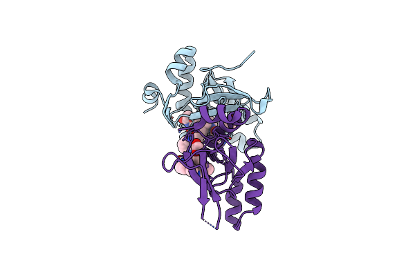 |
Crystal Structure Of The Pwwp1 Domain Of Nsd2 Bound By Compound 17.
Organism: Homo sapiens
Method: X-RAY DIFFRACTION Resolution:2.43 Å Release Date: 2024-05-29 Classification: TRANSFERASE Ligands: EDO, A1H7X |
Organism: Homo sapiens
Method: X-RAY DIFFRACTION
Release Date: 2024-05-29
Ligands: EDO, A1H7X
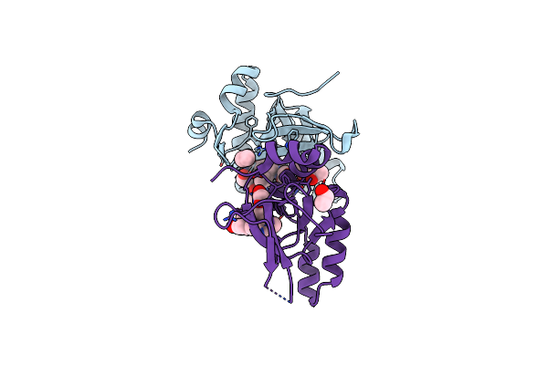 |
Crystal Structure Of The Pwwp1 Domain Of Nsd2 Bound By Compound 18.
Organism: Homo sapiens
Method: X-RAY DIFFRACTION Resolution:1.94 Å Release Date: 2024-05-29 Classification: TRANSFERASE Ligands: EDO, EOH, A1H7Y |
Organism: Homo sapiens
Method: X-RAY DIFFRACTION
Release Date: 2024-05-29
Ligands: EDO, EOH, A1H7Y
 |
Crystal Structure Of The Pwwp1 Domain Of Nsd2 Bound By Compound 34.
Organism: Homo sapiens
Method: X-RAY DIFFRACTION Resolution:1.70 Å Release Date: 2024-05-29 Classification: TRANSFERASE Ligands: A1H7Z |
Organism: Homo sapiens
Method: X-RAY DIFFRACTION
Release Date: 2024-05-29
Ligands: A1H7Z
 |
Structure Of Monobody 27 Human Mlkl Pseudokinase Domain Complex
Organism: Homo sapiens, Synthetic construct
Method: X-RAY DIFFRACTION Resolution:2.63 Å Release Date: 2021-03-10 Classification: TRANSFERASE |
Organism: Homo sapiens, Synthetic construct
Method: X-RAY DIFFRACTION
Release Date: 2021-03-10
 |
Structure Of Monobody 32 Human Mlkl Pseudokinase Domain Complex
Organism: Homo sapiens, Synthetic construct
Method: X-RAY DIFFRACTION Resolution:2.44 Å Release Date: 2021-03-10 Classification: TRANSFERASE Ligands: EDO |
Organism: Homo sapiens, Synthetic construct
Method: X-RAY DIFFRACTION
Release Date: 2021-03-10
Ligands: EDO
 |
Structure Of The Cydia Pomonella Granulovirus Kinase, Pk-1
Organism: Cydia pomonella granulosis virus (isolate mexican)
Method: X-RAY DIFFRACTION Resolution:2.01 Å Release Date: 2021-02-03 Classification: VIRAL PROTEIN Ligands: AMP |
Organism: Cydia pomonella granulosis virus (isolate mexican)
Method: X-RAY DIFFRACTION
Release Date: 2021-02-03
Ligands: AMP
 |
Crystal Structure Of Bak Core Domain Bh3-Groove-Dimer In Complex With E. Coli Lipid
Organism: Homo sapiens
Method: X-RAY DIFFRACTION Resolution:2.49 Å Release Date: 2020-09-02 Classification: APOPTOSIS Ligands: PEE |
Organism: Homo sapiens
Method: X-RAY DIFFRACTION
Release Date: 2020-09-02
Ligands: PEE
 |
Crystal Structure Of Bak Core Domain Bh3-Groove-Dimer In Complex With Phosphatidylserine
Organism: Homo sapiens
Method: X-RAY DIFFRACTION Resolution:2.49 Å Release Date: 2020-09-02 Classification: APOPTOSIS Ligands: 8SP, SO4, GOL |
Organism: Homo sapiens
Method: X-RAY DIFFRACTION
Release Date: 2020-09-02
Ligands: 8SP, SO4, GOL
 |
Crystal Structure Of Bak Core Domain Bh3-Groove-Dimer In Complex With Ddm
Organism: Homo sapiens
Method: X-RAY DIFFRACTION Resolution:1.80 Å Release Date: 2020-09-02 Classification: APOPTOSIS Ligands: LMT, SO4, ACT, EDO |
Organism: Homo sapiens
Method: X-RAY DIFFRACTION
Release Date: 2020-09-02
Ligands: LMT, SO4, ACT, EDO
 |
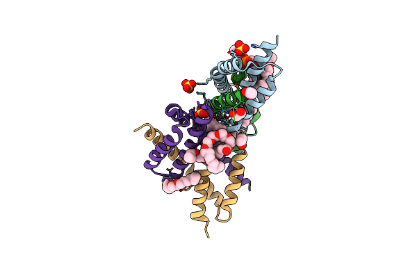
Crystal Structure Of Bak Core Domain Bh3-Groove-Dimer In Complex With Phosphatidylglycerol
Organism: Homo sapiens
Method: X-RAY DIFFRACTION Resolution:2.49 Å Release Date: 2020-09-02 Classification: APOPTOSIS Ligands: PG8, GOL |
Organism: Homo sapiens
Method: X-RAY DIFFRACTION
Release Date: 2020-09-02
Ligands: PG8, GOL
 |
Crystal Structure Of Bak Core Domain Bh3-Groove-Dimer In Complex With Popc And C8E4
Organism: Homo sapiens
Method: X-RAY DIFFRACTION Resolution:1.70 Å Release Date: 2020-09-02 Classification: APOPTOSIS Ligands: SO4, C8E, EDO, LBN |
Organism: Homo sapiens
Method: X-RAY DIFFRACTION
Release Date: 2020-09-02
Ligands: SO4, C8E, EDO, LBN
 |
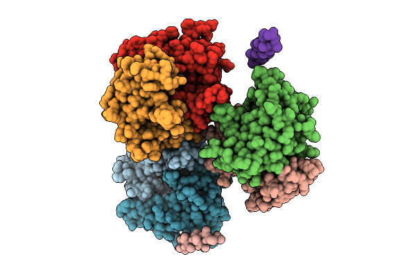
Crystal Structure Of Bak Core Domain Bh3-Groove-Dimer In Complex With Lysopc
Organism: Homo sapiens
Method: X-RAY DIFFRACTION Resolution:1.80 Å Release Date: 2020-09-02 Classification: APOPTOSIS Ligands: K6G, PG4, PGE |
Organism: Homo sapiens
Method: X-RAY DIFFRACTION
Release Date: 2020-09-02
Ligands: K6G, PG4, PGE
 |
Crystal Structure Of Mouse Smchd1 Hinge Domain
Organism: Mus musculus
Method: X-RAY DIFFRACTION Resolution:3.30 Å Release Date: 2020-06-17 Classification: DNA BINDING PROTEIN |
Organism: Mus musculus
Method: X-RAY DIFFRACTION
Release Date: 2020-06-17
 |
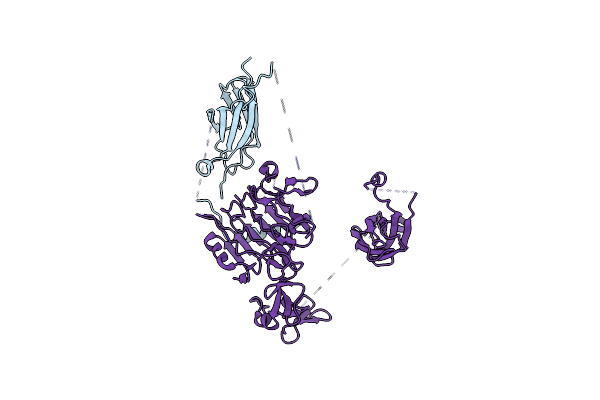
Head Region Of The Open Conformation Of The Human Type 1 Insulin-Like Growth Factor Receptor Ectodomain In Complex With Human Insulin-Like Growth Factor Ii.
Organism: Homo sapiens, Saccharomyces cerevisiae (strain atcc 204508 / s288c)
Method: ELECTRON MICROSCOPY Release Date: 2020-05-13 Classification: SIGNALING PROTEIN Ligands: NAG |
Organism: Homo sapiens, Saccharomyces cerevisiae (strain atcc 204508 / s288c)
Method: ELECTRON MICROSCOPY
Release Date: 2020-05-13
Ligands: NAG
 |
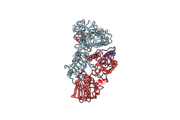
Leg Region Of The Open Conformation Of The Human Type 1 Insulin-Like Growth Factor Receptor Ectodomain In Complex With Human Insulin-Like Growth Factor Ii.
Organism: Homo sapiens, Saccharomyces cerevisiae (strain atcc 204508 / s288c)
Method: ELECTRON MICROSCOPY Release Date: 2020-05-13 Classification: SIGNALING PROTEIN |
Organism: Homo sapiens, Saccharomyces cerevisiae (strain atcc 204508 / s288c)
Method: ELECTRON MICROSCOPY
Release Date: 2020-05-13
 |
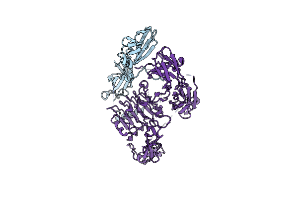
Head Region Of The Closed Conformation Of The Human Type 1 Insulin-Like Growth Factor Receptor Ectodomain In Complex With Human Insulin-Like Growth Factor Ii.
Organism: Homo sapiens, Saccharomyces cerevisiae (strain atcc 204508 / s288c)
Method: ELECTRON MICROSCOPY Release Date: 2020-05-13 Classification: SIGNALING PROTEIN Ligands: NAG |
Organism: Homo sapiens, Saccharomyces cerevisiae (strain atcc 204508 / s288c)
Method: ELECTRON MICROSCOPY
Release Date: 2020-05-13
Ligands: NAG
 |
Leg Region Of The Closed Conformation Of The Human Type 1 Insulin-Like Growth Factor Receptor Ectodomain In Complex With Human Insulin-Like Growth Factor Ii
Organism: Homo sapiens, Saccharomyces cerevisiae (strain atcc 204508 / s288c)
Method: ELECTRON MICROSCOPY Release Date: 2020-05-13 Classification: SIGNALING PROTEIN |
Organism: Homo sapiens, Saccharomyces cerevisiae (strain atcc 204508 / s288c)
Method: ELECTRON MICROSCOPY
Release Date: 2020-05-13
 |
Structure Of Monobody 33 Mlkl N-Terminal Domain Complex
Organism: Homo sapiens, Synthetic construct
Method: X-RAY DIFFRACTION Resolution:2.50 Å Release Date: 2020-04-01 Classification: TRANSFERASE Ligands: ZN |
Organism: Homo sapiens, Synthetic construct
Method: X-RAY DIFFRACTION
Release Date: 2020-04-01
Ligands: ZN