

Search Count: 41
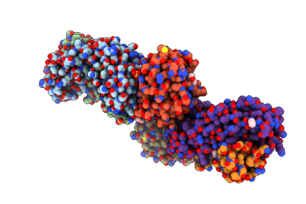 |
Crystal Structure Of Bak In Complex With Inhibiting Antibody 14G6
Organism: Homo sapiens
Method: X-RAY DIFFRACTION Resolution:2.40 Å Release Date: 2024-04-10 Classification: APOPTOSIS/INHIBITOR Ligands: SO4, CCN, PEG, 144, PGE, EDO, 1PE |
Organism: Homo sapiens
Method: X-RAY DIFFRACTION
Release Date: 2024-04-10
Ligands: SO4, CCN, PEG, 144, PGE, EDO, 1PE
 |
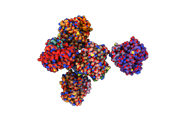
Crystal Structure Of Bax Core Domain Bh3-Groove Dimer - Tetrameric Fraction P21
Organism: Homo sapiens
Method: X-RAY DIFFRACTION Resolution:2.09 Å Release Date: 2023-12-27 Classification: APOPTOSIS Ligands: EDO |
Organism: Homo sapiens
Method: X-RAY DIFFRACTION
Release Date: 2023-12-27
Ligands: EDO
 |
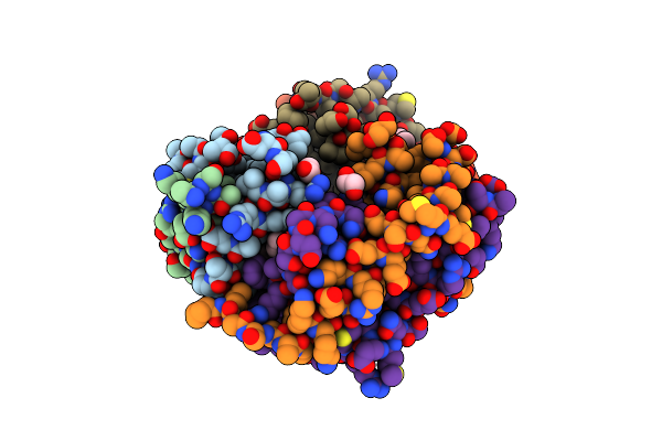
Crystal Structure Of Bax Core Domain Bh3-Groove Dimer - Tetrameric Fraction P31
Organism: Homo sapiens
Method: X-RAY DIFFRACTION Resolution:2.30 Å Release Date: 2023-12-27 Classification: APOPTOSIS Ligands: ZN, EDO, PEG |
Organism: Homo sapiens
Method: X-RAY DIFFRACTION
Release Date: 2023-12-27
Ligands: ZN, EDO, PEG
 |
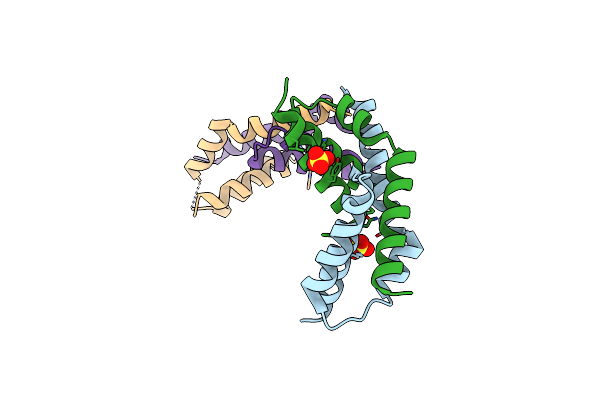
Crystal Structure Of Bax Core Domain Bh3-Groove Dimer - Hexameric Fraction With 2-Stearoyl Lysopc
Organism: Homo sapiens
Method: X-RAY DIFFRACTION Resolution:2.20 Å Release Date: 2023-12-27 Classification: APOPTOSIS Ligands: 1GP, EDO, OCT, D12, DD9 |
Organism: Homo sapiens
Method: X-RAY DIFFRACTION
Release Date: 2023-12-27
Ligands: 1GP, EDO, OCT, D12, DD9
 |
Crystal Structure Of Bax Core Domain Bh3-Groove Dimer - Hexameric Fraction With Dioctanoyl Phosphatidylserine
Organism: Homo sapiens
Method: X-RAY DIFFRACTION Resolution:2.40 Å Release Date: 2023-12-27 Classification: APOPTOSIS Ligands: SO4 |
Organism: Homo sapiens
Method: X-RAY DIFFRACTION
Release Date: 2023-12-27
Ligands: SO4
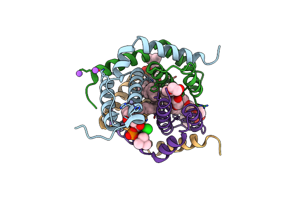 |
Crystal Structure Of Bak-Bax Heterodimer With Lysopc
Organism: Homo sapiens
Method: X-RAY DIFFRACTION Resolution:2.09 Å Release Date: 2023-12-27 Classification: APOPTOSIS Ligands: K6G, ZN, NA, PEG, EDO, CL |
Organism: Homo sapiens
Method: X-RAY DIFFRACTION
Release Date: 2023-12-27
Ligands: K6G, ZN, NA, PEG, EDO, CL
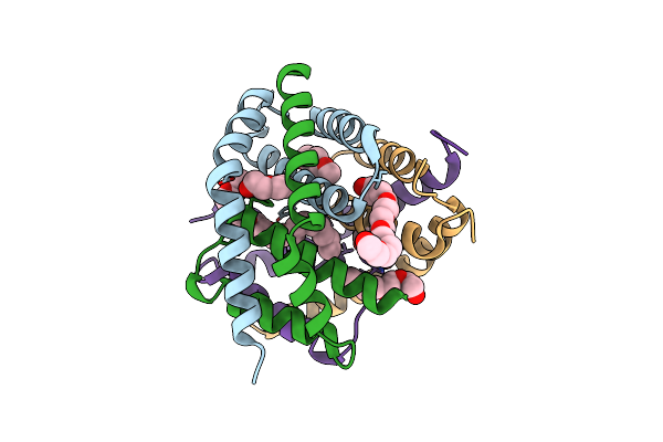 |
Crystal Structure Of Bak-Bax Heterodimer With C12E8
Organism: Homo sapiens
Method: X-RAY DIFFRACTION Resolution:2.40 Å Release Date: 2023-12-27 Classification: APOPTOSIS Ligands: PG0, PEG, N8E, P33, PG4 |
Organism: Homo sapiens
Method: X-RAY DIFFRACTION
Release Date: 2023-12-27
Ligands: PG0, PEG, N8E, P33, PG4
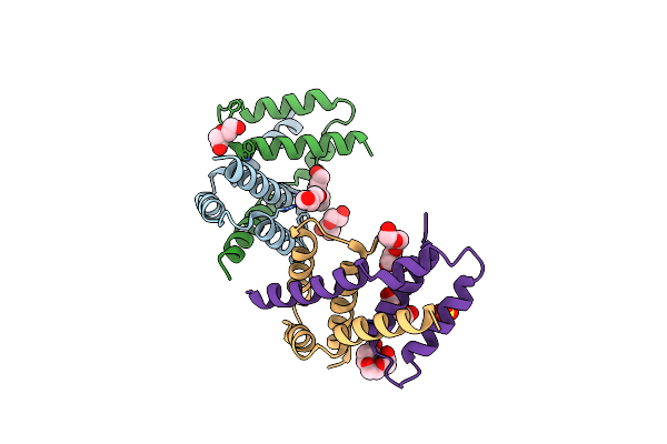 |
Crystal Structure Of Bax D71N Core Domain Bh3-Groove Dimer
Organism: Homo sapiens
Method: X-RAY DIFFRACTION Resolution:2.25 Å Release Date: 2023-12-27 Classification: APOPTOSIS Ligands: PEG, PGE, PG4, SO4 |
Organism: Homo sapiens
Method: X-RAY DIFFRACTION
Release Date: 2023-12-27
Ligands: PEG, PGE, PG4, SO4
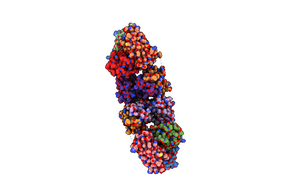 |
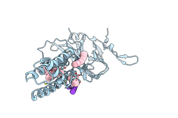
Crystal Structure Of Bak L100A In Complex With Activating Antibody Fragments
Organism: Homo sapiens, Rattus norvegicus
Method: X-RAY DIFFRACTION Resolution:3.10 Å Release Date: 2022-02-09 Classification: APOPTOSIS Ligands: MPD |
Organism: Homo sapiens, Rattus norvegicus
Method: X-RAY DIFFRACTION
Release Date: 2022-02-09
Ligands: MPD
 |
Kirbac3.1 L124M Mutant
Organism: Magnetospirillum magnetotacticum
Method: X-RAY DIFFRACTION Resolution:2.72 Å Release Date: 2021-12-29 Classification: MEMBRANE PROTEIN Ligands: PCW, TMO, OCT, K |
Organism: Magnetospirillum magnetotacticum
Method: X-RAY DIFFRACTION
Release Date: 2021-12-29
Ligands: PCW, TMO, OCT, K
 |
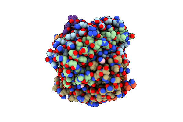
Kirbac3.1 C71S C262S
Organism: Magnetospirillum magnetotacticum
Method: X-RAY DIFFRACTION Resolution:2.40 Å Release Date: 2021-12-29 Classification: MEMBRANE PROTEIN Ligands: PCW, TMO, K |
Organism: Magnetospirillum magnetotacticum
Method: X-RAY DIFFRACTION
Release Date: 2021-12-29
Ligands: PCW, TMO, K
 |
Crystal Structure Of Bak Core Domain Bh3-Groove-Dimer In Complex With E. Coli Lipid
Organism: Homo sapiens
Method: X-RAY DIFFRACTION Resolution:2.49 Å Release Date: 2020-09-02 Classification: APOPTOSIS Ligands: PEE |
Organism: Homo sapiens
Method: X-RAY DIFFRACTION
Release Date: 2020-09-02
Ligands: PEE
 |
Crystal Structure Of Bak Core Domain Bh3-Groove-Dimer In Complex With Phosphatidylserine
Organism: Homo sapiens
Method: X-RAY DIFFRACTION Resolution:2.49 Å Release Date: 2020-09-02 Classification: APOPTOSIS Ligands: 8SP, SO4, GOL |
Organism: Homo sapiens
Method: X-RAY DIFFRACTION
Release Date: 2020-09-02
Ligands: 8SP, SO4, GOL
 |
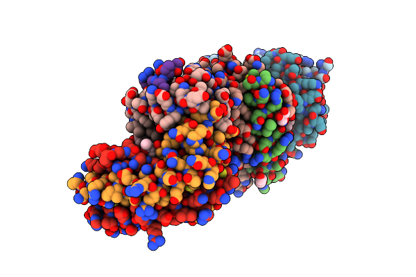
Crystal Structure Of Bak Core Domain Bh3-Groove-Dimer In Complex With Ddm
Organism: Homo sapiens
Method: X-RAY DIFFRACTION Resolution:1.80 Å Release Date: 2020-09-02 Classification: APOPTOSIS Ligands: LMT, SO4, ACT, EDO |
Organism: Homo sapiens
Method: X-RAY DIFFRACTION
Release Date: 2020-09-02
Ligands: LMT, SO4, ACT, EDO
 |
Crystal Structure Of Bak Core Domain Bh3-Groove-Dimer In Complex With Phosphatidylglycerol
Organism: Homo sapiens
Method: X-RAY DIFFRACTION Resolution:2.49 Å Release Date: 2020-09-02 Classification: APOPTOSIS Ligands: PG8, GOL |
Organism: Homo sapiens
Method: X-RAY DIFFRACTION
Release Date: 2020-09-02
Ligands: PG8, GOL
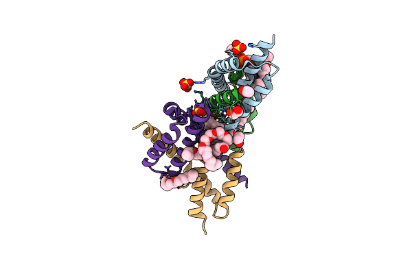 |
Crystal Structure Of Bak Core Domain Bh3-Groove-Dimer In Complex With Popc And C8E4
Organism: Homo sapiens
Method: X-RAY DIFFRACTION Resolution:1.70 Å Release Date: 2020-09-02 Classification: APOPTOSIS Ligands: SO4, C8E, EDO, LBN |
Organism: Homo sapiens
Method: X-RAY DIFFRACTION
Release Date: 2020-09-02
Ligands: SO4, C8E, EDO, LBN
 |
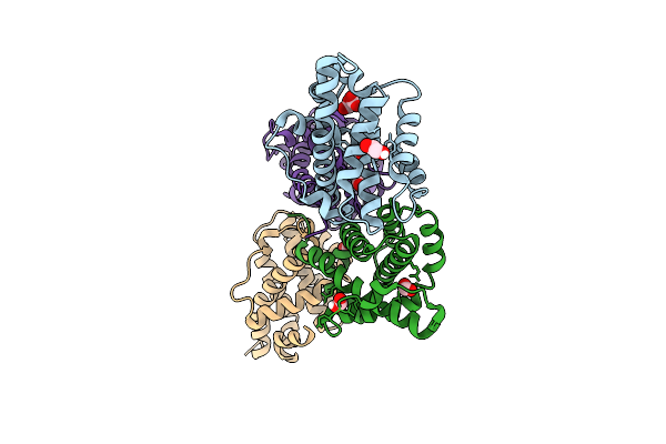
Crystal Structure Of Bak Core Domain Bh3-Groove-Dimer In Complex With Lysopc
Organism: Homo sapiens
Method: X-RAY DIFFRACTION Resolution:1.80 Å Release Date: 2020-09-02 Classification: APOPTOSIS Ligands: K6G, PG4, PGE |
Organism: Homo sapiens
Method: X-RAY DIFFRACTION
Release Date: 2020-09-02
Ligands: K6G, PG4, PGE
 |
Crystal Structure Of Mouse Bak
Organism: Mus musculus
Method: X-RAY DIFFRACTION Resolution:1.75 Å Release Date: 2019-09-11 Classification: APOPTOSIS Ligands: FMT, EDO |
Organism: Mus musculus
Method: X-RAY DIFFRACTION
Release Date: 2019-09-11
Ligands: FMT, EDO
 |
Crystal Structure Of Baxp168G In Complex With An Activating Antibody
Organism: Homo sapiens, Rattus norvegicus
Method: X-RAY DIFFRACTION Resolution:2.50 Å Release Date: 2018-06-27 Classification: APOPTOSIS/IMMUNE SYSTEM Ligands: EDO, ZN |
Organism: Homo sapiens, Rattus norvegicus
Method: X-RAY DIFFRACTION
Release Date: 2018-06-27
Ligands: EDO, ZN
 |
Crystal Structure Of Baxp168G In Complex With An Activating Antibody At High Resolution
Organism: Homo sapiens, Rattus norvegicus
Method: X-RAY DIFFRACTION Resolution:2.00 Å Release Date: 2018-06-27 Classification: APOPTOSIS Ligands: EDO, PG4, PGE, MES |
Organism: Homo sapiens, Rattus norvegicus
Method: X-RAY DIFFRACTION
Release Date: 2018-06-27
Ligands: EDO, PG4, PGE, MES