

Search Count: 818
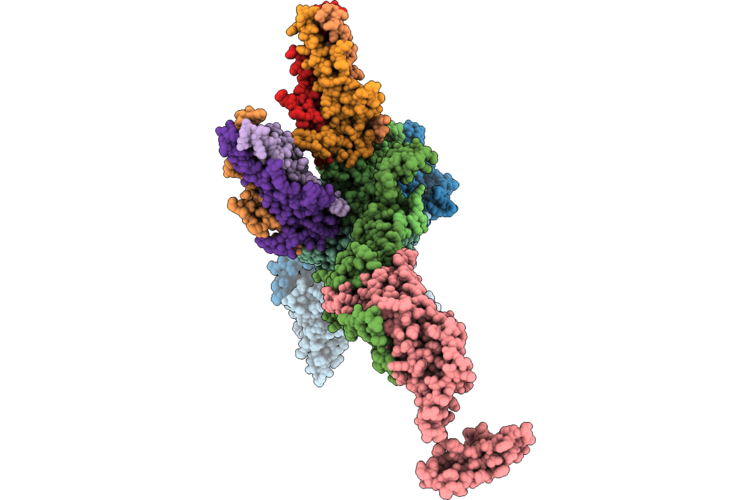 |
Phage T4 Neck In Post-Tail-Contraction State (Genome-Full Particle)
Organism: Escherichia phage t4
Method: ELECTRON MICROSCOPY Release Date: 2026-07-22 Classification: VIRAL PROTEIN |
Organism: Escherichia phage t4
Method: ELECTRON MICROSCOPY
Release Date: 2026-07-22
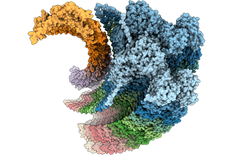 |
Phage T4 Sheath In Post-Tail-Contraction State (Genome-Full Particle)
Organism: Escherichia phage t4
Method: ELECTRON MICROSCOPY Release Date: 2026-07-22 Classification: VIRAL PROTEIN |
Organism: Escherichia phage t4
Method: ELECTRON MICROSCOPY
Release Date: 2026-07-22
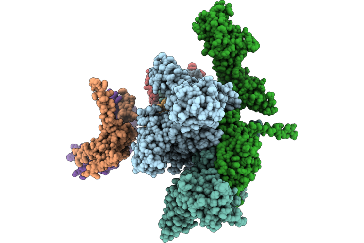 |
Phage T4 Inner Baseplate In Post-Tail-Contraction State (Genome-Full Particle)
Organism: Escherichia phage t4
Method: ELECTRON MICROSCOPY Release Date: 2026-07-22 Classification: VIRAL PROTEIN |
Organism: Escherichia phage t4
Method: ELECTRON MICROSCOPY
Release Date: 2026-07-22
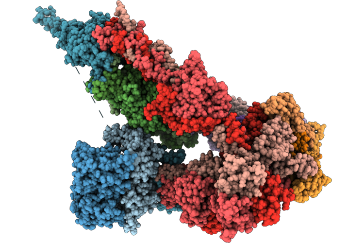 |
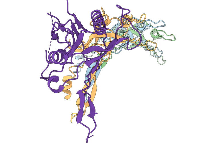
Phage T4 Peripheral Baseplate In Post-Tail-Contraction State (Genome-Full Particle)
Organism: Escherichia phage t4
Method: ELECTRON MICROSCOPY Release Date: 2026-07-22 Classification: VIRAL PROTEIN |
Organism: Escherichia phage t4
Method: ELECTRON MICROSCOPY
Release Date: 2026-07-22
 |
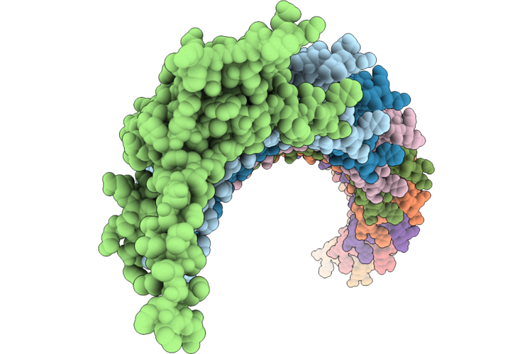
Phage T4 Tip Of The Tail Tube In Post-Tail-Contraction State (Genome-Empty Particle)
Organism: Escherichia phage t4
Method: ELECTRON MICROSCOPY Release Date: 2026-07-22 Classification: VIRAL PROTEIN |
Organism: Escherichia phage t4
Method: ELECTRON MICROSCOPY
Release Date: 2026-07-22
 |
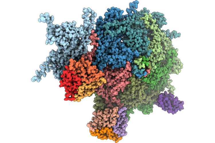
Phage T4 Protruding Part Of The Tail Tube In Post-Tail-Contraction State (Genome-Full Particle)
Organism: Escherichia phage t4
Method: ELECTRON MICROSCOPY Resolution:3.54 Å Release Date: 2026-07-22 Classification: VIRAL PROTEIN |
Organism: Escherichia phage t4
Method: ELECTRON MICROSCOPY
Release Date: 2026-07-22
 |
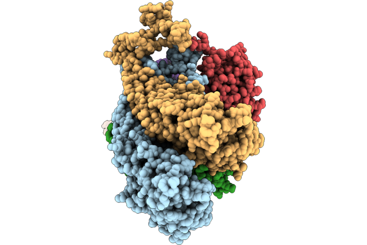
Cryo-Em Structure Of Parabacteroide Phage Pd491P1 Capsid
Organism: Parabacteroides phage pd491p1
Method: ELECTRON MICROSCOPY Release Date: 2026-06-24 Classification: VIRUS |
Organism: Parabacteroides phage pd491p1
Method: ELECTRON MICROSCOPY
Release Date: 2026-06-24
 |
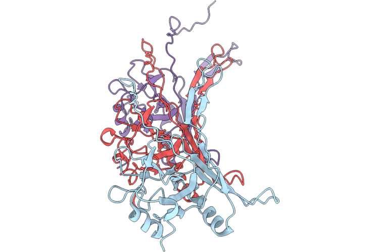
Cryo-Em Structure Of Parabacteroide Phage Pd491P1 Head-To-Tail Interface
Organism: Parabacteroides phage pd491p1
Method: ELECTRON MICROSCOPY Release Date: 2026-06-24 Classification: VIRAL PROTEIN |
Organism: Parabacteroides phage pd491p1
Method: ELECTRON MICROSCOPY
Release Date: 2026-06-24
 |
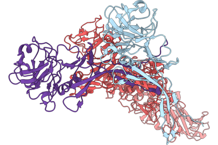
Cryo-Em Structure Of The Tail Tip Region Of Parabacteroide Phage Pd491P1 (Imposed With C6 Symmetry)
Organism: Parabacteroides phage pd491p1
Method: ELECTRON MICROSCOPY Release Date: 2026-06-24 Classification: VIRAL PROTEIN |
Organism: Parabacteroides phage pd491p1
Method: ELECTRON MICROSCOPY
Release Date: 2026-06-24
 |
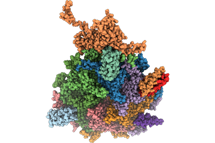
Cryo-Em Structure Of The Tail Tip Region Of Parabacteroide Phage Pd491P1 (Imposed With C3 Symmetry)
Organism: Parabacteroides phage pd491p1
Method: ELECTRON MICROSCOPY Release Date: 2026-06-24 Classification: VIRAL PROTEIN |
Organism: Parabacteroides phage pd491p1
Method: ELECTRON MICROSCOPY
Release Date: 2026-06-24
 |
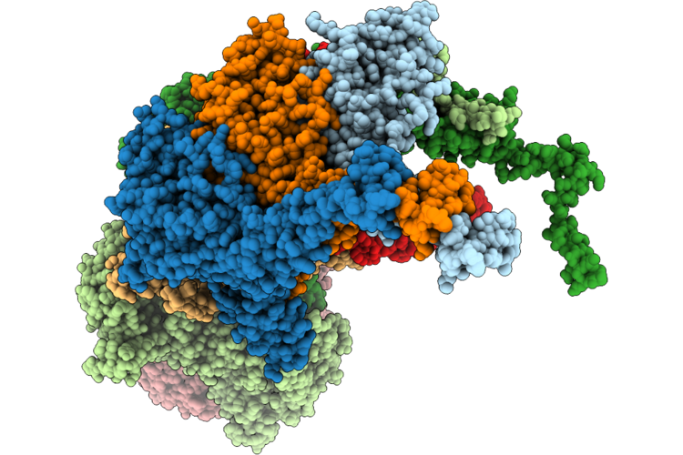
Cryo-Em Structure Of Icosahedrally Averaged Bacteriophage Ran69 Capsid
Organism: Klebsiella phage ran69
Method: ELECTRON MICROSCOPY Release Date: 2026-05-27 Classification: VIRUS |
Organism: Klebsiella phage ran69
Method: ELECTRON MICROSCOPY
Release Date: 2026-05-27
 |
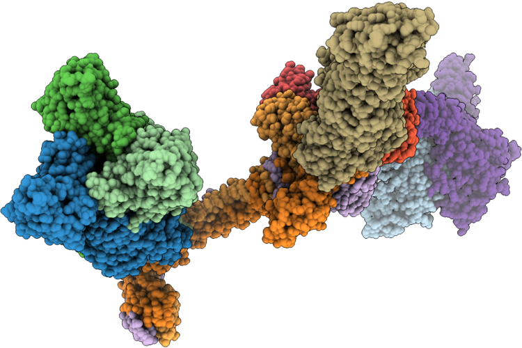
The Composite Cryo-Em Structure Of Bacteriophage Ran69 Pre-Ejectosome-Portal Complex
Organism: Klebsiella phage ran69
Method: ELECTRON MICROSCOPY Release Date: 2026-05-27 Classification: VIRUS |
Organism: Klebsiella phage ran69
Method: ELECTRON MICROSCOPY
Release Date: 2026-05-27
 |
The Composite Cryo-Em Structure Of The Tail Region Of Bacteriophage Ran69
Organism: Klebsiella phage ran69
Method: ELECTRON MICROSCOPY Resolution:3.20 Å Release Date: 2026-05-27 Classification: VIRUS |
Organism: Klebsiella phage ran69
Method: ELECTRON MICROSCOPY
Release Date: 2026-05-27
 |
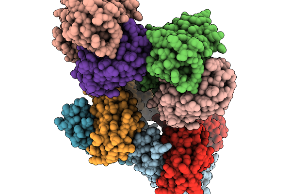
Phage T4 Tip Of The Tail Tube In Post-Tail-Contraction State (Genome-Full Particle)
Organism: Escherichia phage t4
Method: ELECTRON MICROSCOPY Release Date: 2026-04-15 Classification: VIRAL PROTEIN |
Organism: Escherichia phage t4
Method: ELECTRON MICROSCOPY
Release Date: 2026-04-15
 |
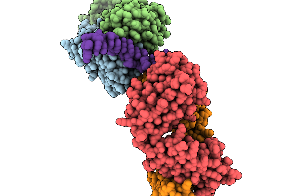
Crystal Structure Of Chimeric Anti-Z-Dna Fab Cz22-Fab
Organism: Mus musculus
Method: X-RAY DIFFRACTION Resolution:2.15 Å Release Date: 2026-02-18 Classification: IMMUNE SYSTEM |
Organism: Mus musculus
Method: X-RAY DIFFRACTION
Release Date: 2026-02-18
 |
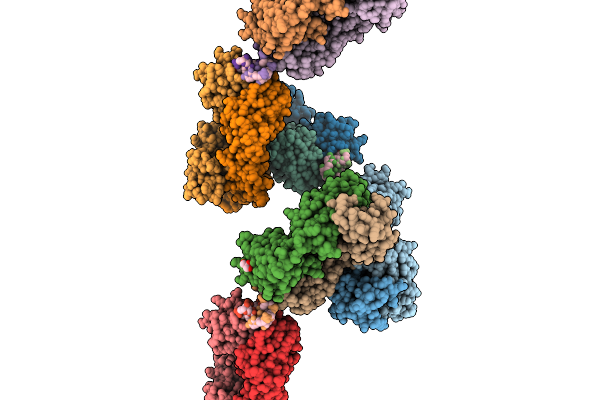
Crystal Structure Of Cz22-Fab In Complex With Left-Handed D(Cg)6 Dna
Organism: Mus musculus, Homo sapiens
Method: X-RAY DIFFRACTION Resolution:3.27 Å Release Date: 2026-02-18 Classification: IMMUNE SYSTEM |
Organism: Mus musculus, Homo sapiens
Method: X-RAY DIFFRACTION
Release Date: 2026-02-18
 |
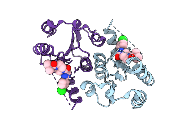
Crystal Structure Of Cz22-Fab In Complex With Left-Handed Dc(Gc)3 Dna
Organism: Mus musculus, Homo sapiens
Method: X-RAY DIFFRACTION Resolution:2.35 Å Release Date: 2026-02-18 Classification: IMMUNE SYSTEM Ligands: GOL |
Organism: Mus musculus, Homo sapiens
Method: X-RAY DIFFRACTION
Release Date: 2026-02-18
Ligands: GOL
 |
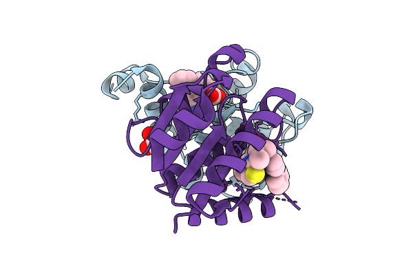
Crystal Structure Of Dnph1 Bound By Compound 3
Organism: Homo sapiens
Method: X-RAY DIFFRACTION Resolution:2.02 Å Release Date: 2025-12-24 Classification: HYDROLASE Ligands: A1JH5 |
Organism: Homo sapiens
Method: X-RAY DIFFRACTION
Release Date: 2025-12-24
Ligands: A1JH5
 |
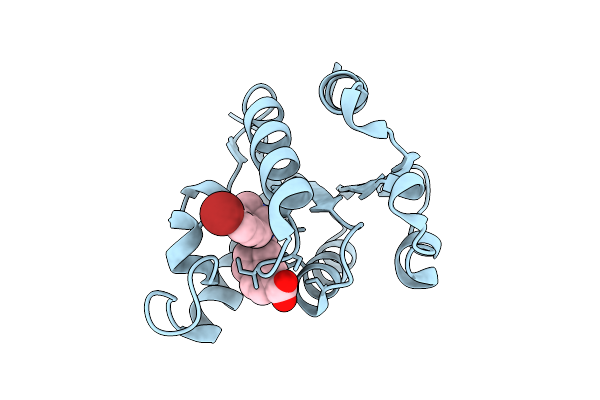
Crystal Structure Of Dnph1 Bound By Compound 6
Organism: Homo sapiens
Method: X-RAY DIFFRACTION Resolution:1.51 Å Release Date: 2025-12-24 Classification: HYDROLASE Ligands: A1JKT, EDO |
Organism: Homo sapiens
Method: X-RAY DIFFRACTION
Release Date: 2025-12-24
Ligands: A1JKT, EDO
 |
Crystal Structure Of Dnph1 Bound By Compound 19
Organism: Homo sapiens
Method: X-RAY DIFFRACTION Resolution:1.50 Å Release Date: 2025-12-24 Classification: HYDROLASE Ligands: A1JKZ |
Organism: Homo sapiens
Method: X-RAY DIFFRACTION
Release Date: 2025-12-24
Ligands: A1JKZ