

Search Count: 34
 |
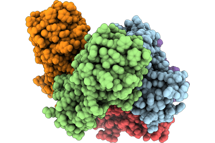
Structure Of E. Roggenkampii Caps
Organism: Enterobacter roggenkampii
Method: X-RAY DIFFRACTION Resolution:1.55 Å Release Date: 2026-06-03 Classification: DNA BINDING PROTEIN Ligands: SO4 |
Organism: Enterobacter roggenkampii
Method: X-RAY DIFFRACTION
Release Date: 2026-06-03
Ligands: SO4
 |
Structure Of V. Cholerae Caps (Form 1)
Organism: Vibrio cholerae
Method: X-RAY DIFFRACTION Resolution:2.38 Å Release Date: 2026-06-03 Classification: DNA BINDING PROTEIN |
Organism: Vibrio cholerae
Method: X-RAY DIFFRACTION
Release Date: 2026-06-03
 |
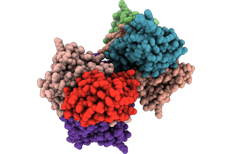
Structure Of V. Cholerae Caps (Form 2)
Organism: Vibrio cholerae
Method: X-RAY DIFFRACTION Resolution:1.84 Å Release Date: 2026-06-03 Classification: DNA BINDING PROTEIN Ligands: SO4 |
Organism: Vibrio cholerae
Method: X-RAY DIFFRACTION
Release Date: 2026-06-03
Ligands: SO4
 |
Structure Of Escherichia Vaps-Vapc Complex
Organism: Escherichia
Method: X-RAY DIFFRACTION Resolution:3.40 Å Release Date: 2026-06-03 Classification: TOXIN Ligands: MG |
Organism: Escherichia
Method: X-RAY DIFFRACTION
Release Date: 2026-06-03
Ligands: MG
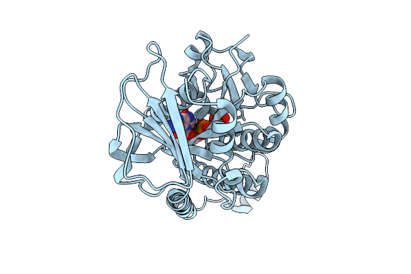 |
Phuz Tubulin From Phage Goslar (Locally Refined Monomer)
Organism: Goslarvirus
Method: ELECTRON MICROSCOPY Resolution:2.75 Å Release Date: 2025-12-24 Classification: VIRAL PROTEIN Ligands: G2P |
Organism: Goslarvirus
Method: ELECTRON MICROSCOPY
Release Date: 2025-12-24
Ligands: G2P
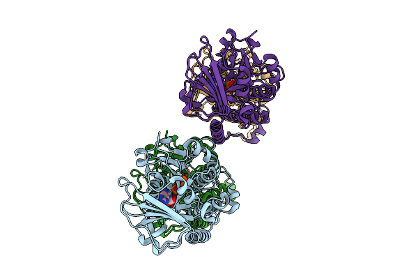 |
Phuz Tubulin Tetramer From Phage Goslar
Organism: Goslarvirus
Method: ELECTRON MICROSCOPY Resolution:2.84 Å Release Date: 2025-12-24 Classification: VIRAL PROTEIN Ligands: G2P |
Organism: Goslarvirus
Method: ELECTRON MICROSCOPY
Release Date: 2025-12-24
Ligands: G2P
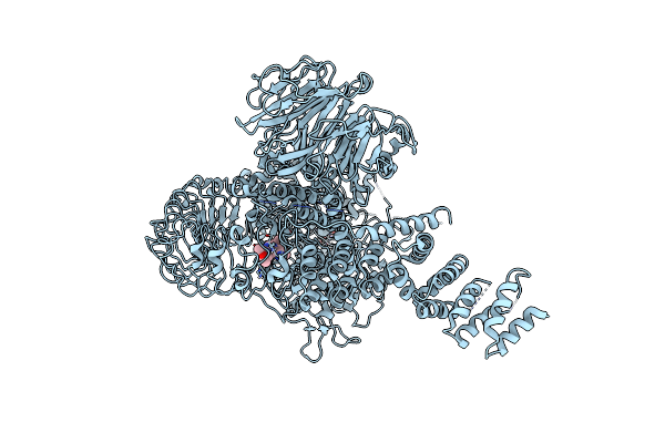 |
Autoinhibited Full-Length Lrrk2(I2020T) On Microtubules With Mli-2
Organism: Homo sapiens
Method: ELECTRON MICROSCOPY Release Date: 2024-12-25 Classification: PROTEIN BINDING Ligands: GDP, A1N |
Organism: Homo sapiens
Method: ELECTRON MICROSCOPY
Release Date: 2024-12-25
Ligands: GDP, A1N
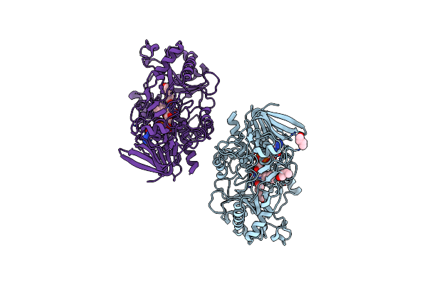 |
Crystal Structure Of An Alcohol-Oxidase From Sphingobacterium Daejeonense
Organism: Sphingobacterium daejeonense
Method: X-RAY DIFFRACTION Resolution:2.00 Å Release Date: 2024-11-13 Classification: OXIDOREDUCTASE Ligands: 1BO, FAD, HEZ |
Organism: Sphingobacterium daejeonense
Method: X-RAY DIFFRACTION
Release Date: 2024-11-13
Ligands: 1BO, FAD, HEZ
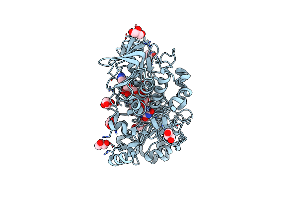 |
Crystal Structure Of An Alcohol Oxidase From Streptomyces Hiroshimensis
Organism: Streptomyces hiroshimensis
Method: X-RAY DIFFRACTION Resolution:2.00 Å Release Date: 2024-11-13 Classification: OXIDOREDUCTASE Ligands: FAD, PGE, GOL |
Organism: Streptomyces hiroshimensis
Method: X-RAY DIFFRACTION
Release Date: 2024-11-13
Ligands: FAD, PGE, GOL
 |
Structure Of Bacteriophage Pa1C Gp2
Organism: Pseudomonas phage pa1c
Method: X-RAY DIFFRACTION Resolution:2.63 Å Release Date: 2023-05-10 Classification: VIRAL PROTEIN |
Organism: Pseudomonas phage pa1c
Method: X-RAY DIFFRACTION
Release Date: 2023-05-10
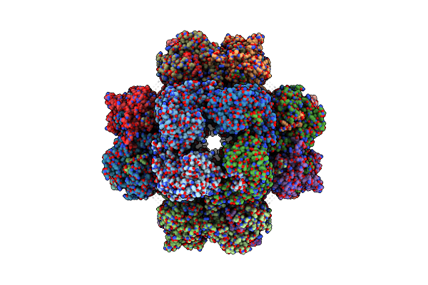 |
201Phi2-1 Chimallin Cubic (O, 24Mer) Assembly
Organism: Pseudomonas phage 201phi2-1
Method: ELECTRON MICROSCOPY Release Date: 2022-07-27 Classification: STRUCTURAL PROTEIN |
Organism: Pseudomonas phage 201phi2-1
Method: ELECTRON MICROSCOPY
Release Date: 2022-07-27
 |
201Phi2-1 Chimallin Localized Tetramer Reconstruction
Organism: Pseudomonas phage 201phi2-1
Method: ELECTRON MICROSCOPY Release Date: 2022-07-27 Classification: STRUCTURAL PROTEIN |
Organism: Pseudomonas phage 201phi2-1
Method: ELECTRON MICROSCOPY
Release Date: 2022-07-27
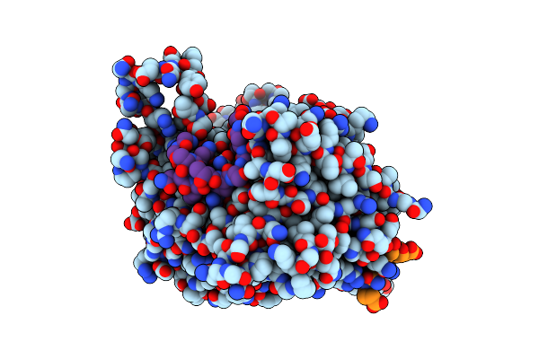 |
201Phi2-1 Chimallin C1 Localized Reconstruction
Organism: Pseudomonas phage 201phi2-1
Method: ELECTRON MICROSCOPY Release Date: 2022-07-27 Classification: STRUCTURAL PROTEIN |
Organism: Pseudomonas phage 201phi2-1
Method: ELECTRON MICROSCOPY
Release Date: 2022-07-27
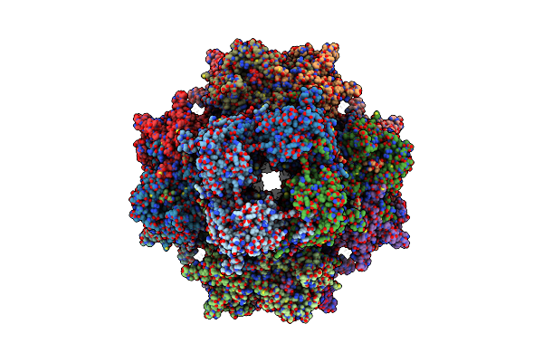 |
Goslar Chimallin Cubic (O, 24Mer) Assembly
Organism: Escherichia phage vb_ecom_goslar
Method: ELECTRON MICROSCOPY Release Date: 2022-07-27 Classification: STRUCTURAL PROTEIN |
Organism: Escherichia phage vb_ecom_goslar
Method: ELECTRON MICROSCOPY
Release Date: 2022-07-27
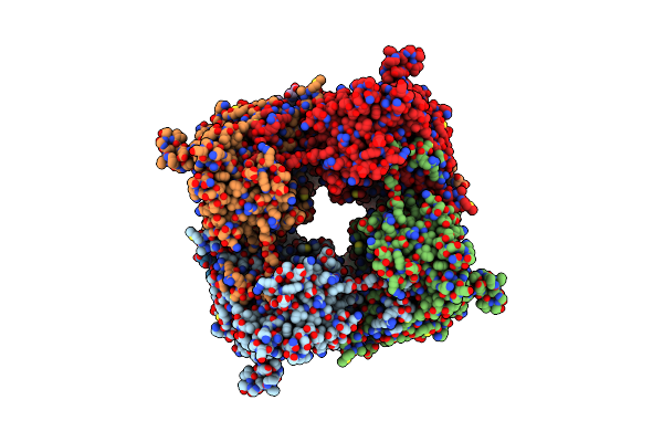 |
Goslar Chimallin C4 Tetramer Localized Reconstruction
Organism: Escherichia phage vb_ecom_goslar
Method: ELECTRON MICROSCOPY Release Date: 2022-07-27 Classification: STRUCTURAL PROTEIN |
Organism: Escherichia phage vb_ecom_goslar
Method: ELECTRON MICROSCOPY
Release Date: 2022-07-27
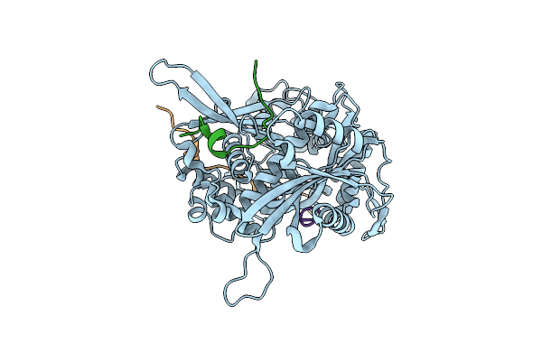 |
Goslar Chimallin C1 Localized Reconstruction
Organism: Escherichia phage vb_ecom_goslar
Method: ELECTRON MICROSCOPY Release Date: 2022-07-27 Classification: STRUCTURAL PROTEIN |
Organism: Escherichia phage vb_ecom_goslar
Method: ELECTRON MICROSCOPY
Release Date: 2022-07-27
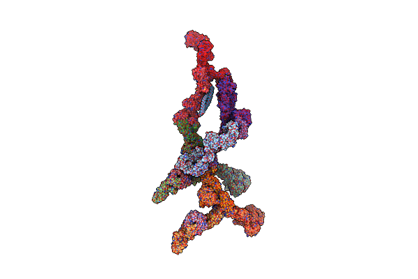 |
Double Nuclear Outer Ring From The Isolated Yeast Npc
Organism: Saccharomyces cerevisiae
Method: ELECTRON MICROSCOPY Release Date: 2022-01-26 Classification: TRANSLOCASE |
Organism: Saccharomyces cerevisiae
Method: ELECTRON MICROSCOPY
Release Date: 2022-01-26
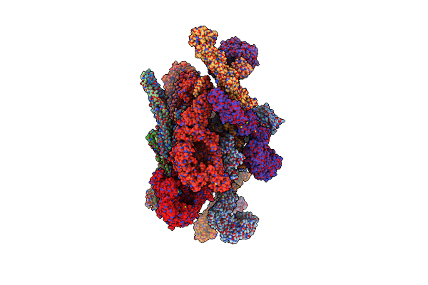 |
Inner Ring Spoke From The Isolated Yeast Npc
Organism: Saccharomyces cerevisiae
Method: ELECTRON MICROSCOPY Release Date: 2022-01-26 Classification: TRANSLOCASE |
Organism: Saccharomyces cerevisiae
Method: ELECTRON MICROSCOPY
Release Date: 2022-01-26
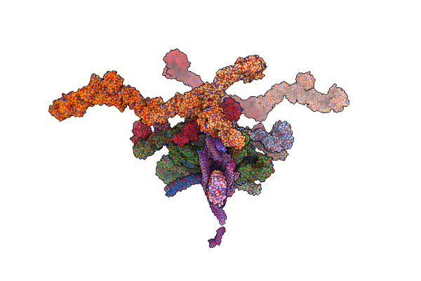 |
Structure Of The In Situ Yeast Npc
Organism: Saccharomyces cerevisiae
Method: ELECTRON MICROSCOPY Release Date: 2022-01-26 Classification: TRANSLOCASE |
Organism: Saccharomyces cerevisiae
Method: ELECTRON MICROSCOPY
Release Date: 2022-01-26
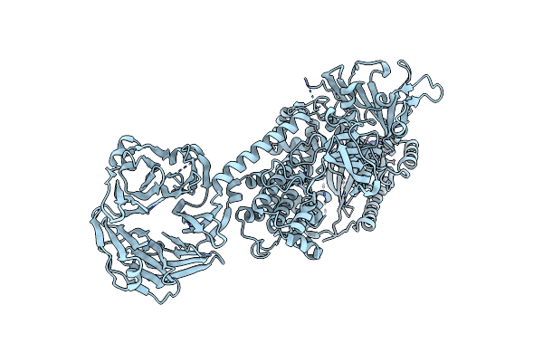 |
Cryo-Em Structure Of The C-Terminal Half Of The Parkinson'S Disease-Linked Protein Leucine Rich Repeat Kinase 2 (Lrrk2)
Organism: Homo sapiens
Method: ELECTRON MICROSCOPY Release Date: 2020-08-26 Classification: SIGNALING PROTEIN Ligands: GDP, MG |
Organism: Homo sapiens
Method: ELECTRON MICROSCOPY
Release Date: 2020-08-26
Ligands: GDP, MG