

Search Count: 21
 |
Crystal Structure Of Xanthobacter Autotrophicus Sparda Mutant Lacking Dren Nuclease Domains
Organism: Xanthobacter autotrophicus py2
Method: X-RAY DIFFRACTION Resolution:1.50 Å Release Date: 2025-12-17 Classification: RNA BINDING PROTEIN Ligands: ACY, GOL, TRS, PEG, CL, BME, TAR Modified Residue(s): MSE, CME |
Organism: Xanthobacter autotrophicus py2
Method: X-RAY DIFFRACTION
Release Date: 2025-12-17
Ligands: ACY, GOL, TRS, PEG, CL, BME, TAR
 |
Crystal Structure Of Enhydrobacter Aerosaccus Apo Sparda Complex
Organism: Enhydrobacter aerosaccus
Method: X-RAY DIFFRACTION Resolution:2.45 Å Release Date: 2025-12-17 Classification: RNA BINDING PROTEIN Ligands: CL, TRS, BME |
Organism: Enhydrobacter aerosaccus
Method: X-RAY DIFFRACTION
Release Date: 2025-12-17
Ligands: CL, TRS, BME
 |
Crystal Structure Of Apo Sparda Complex From Xanthobacter Autotrophicus
Organism: Xanthobacter autotrophicus py2
Method: X-RAY DIFFRACTION Resolution:3.00 Å Release Date: 2025-12-17 Classification: RNA BINDING PROTEIN Ligands: SO4 |
Organism: Xanthobacter autotrophicus py2
Method: X-RAY DIFFRACTION
Release Date: 2025-12-17
Ligands: SO4
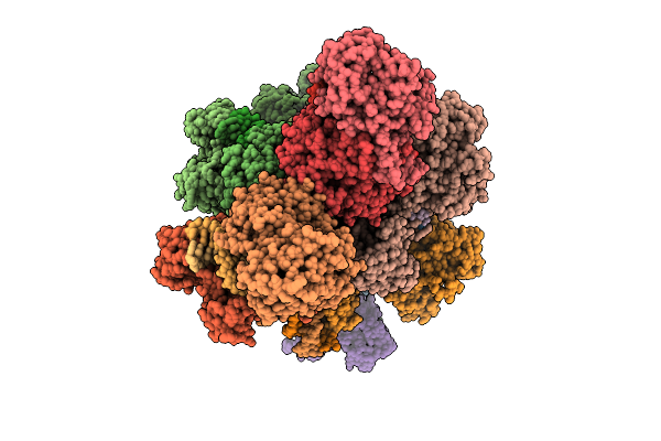 |
Activated Xausparda Filament Assembly With Bound Dsdna Substrate
Organism: Xanthobacter autotrophicus py2, Escherichia coli
Method: ELECTRON MICROSCOPY Release Date: 2025-12-17 Classification: RNA BINDING PROTEIN Ligands: CA |
Organism: Xanthobacter autotrophicus py2, Escherichia coli
Method: ELECTRON MICROSCOPY
Release Date: 2025-12-17
Ligands: CA
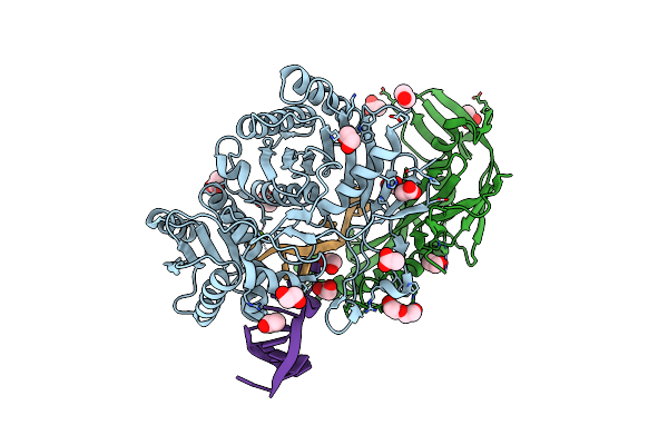 |
Heterodimeric Complex Of Archaeoglobus Fulgidus Argonaute Protein Af1318 (Afago) With Dna And Afago-N Protein Containing N-L1-L2 Domains
Organism: Archaeoglobus fulgidus dsm 4304, Escherichia coli
Method: X-RAY DIFFRACTION Resolution:2.50 Å Release Date: 2024-01-24 Classification: RNA BINDING PROTEIN Ligands: MG, ACY, EDO, PEG |
Organism: Archaeoglobus fulgidus dsm 4304, Escherichia coli
Method: X-RAY DIFFRACTION
Release Date: 2024-01-24
Ligands: MG, ACY, EDO, PEG
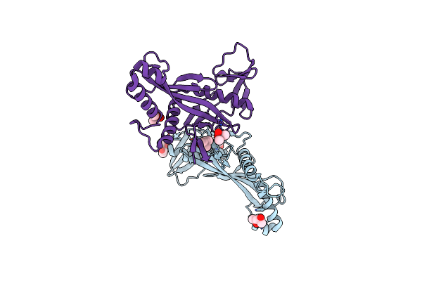 |
Crystal Structure Of Archaeoglobus Fulgidus Afago-N Protein Representing N-L1-L2 Domains
Organism: Archaeoglobus fulgidus dsm 8774
Method: X-RAY DIFFRACTION Resolution:1.85 Å Release Date: 2024-01-24 Classification: RNA BINDING PROTEIN Ligands: CAC, CL, MPD, NA |
Organism: Archaeoglobus fulgidus dsm 8774
Method: X-RAY DIFFRACTION
Release Date: 2024-01-24
Ligands: CAC, CL, MPD, NA
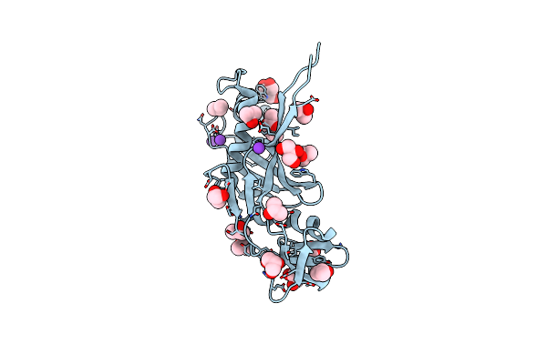 |
Crystal Structure Of Archaeoglobus Fulgidus Afago-N Protein Representing N-L1-L2 Domains
Organism: Archaeoglobus fulgidus dsm 8774
Method: X-RAY DIFFRACTION Resolution:1.40 Å Release Date: 2024-01-24 Classification: RNA BINDING PROTEIN Ligands: GOL, IPA, K, TRS, ACY |
Organism: Archaeoglobus fulgidus dsm 8774
Method: X-RAY DIFFRACTION
Release Date: 2024-01-24
Ligands: GOL, IPA, K, TRS, ACY
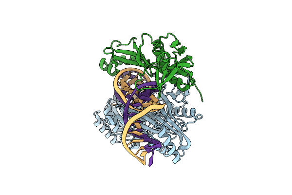 |
Archaeoglobus Fulgidus Afago Complex With Afago-N Protein (Fafago) Bound With 30 Nt Rna Guide And 51 Nt Dna Target
Organism: Archaeoglobus fulgidus, Archaeoglobus fulgidus dsm 8774, Escherichia coli
Method: ELECTRON MICROSCOPY Release Date: 2024-01-24 Classification: DNA BINDING PROTEIN Ligands: MG |
Organism: Archaeoglobus fulgidus, Archaeoglobus fulgidus dsm 8774, Escherichia coli
Method: ELECTRON MICROSCOPY
Release Date: 2024-01-24
Ligands: MG
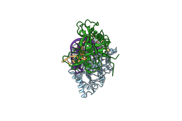 |
Archaeoglobus Fulgidus Afago Complex With Afago-N Protein (Fafago) Bound With 17 Nt Rna Guide And 17 Nt Dna Target
Organism: Archaeoglobus fulgidus, Escherichia coli
Method: ELECTRON MICROSCOPY Release Date: 2024-01-24 Classification: DNA BINDING PROTEIN |
Organism: Archaeoglobus fulgidus, Escherichia coli
Method: ELECTRON MICROSCOPY
Release Date: 2024-01-24
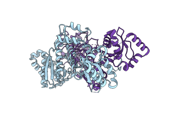 |
Crystal Structure Of Apo Cami1
Organism: Allochromatium vinosum
Method: X-RAY DIFFRACTION Resolution:1.70 Å Release Date: 2023-12-13 Classification: HYDROLASE |
Organism: Allochromatium vinosum
Method: X-RAY DIFFRACTION
Release Date: 2023-12-13
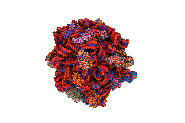 |
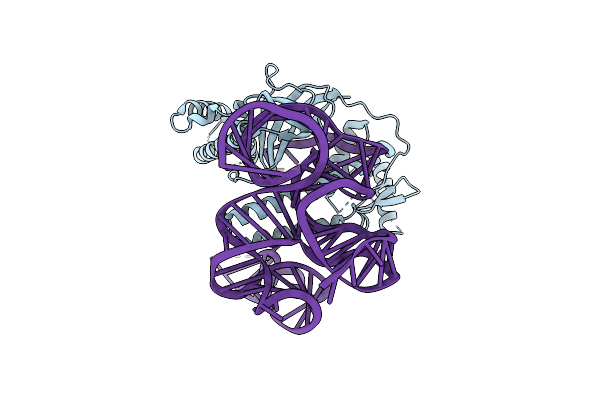
Ca4-Bound Cami1 In Complex With 70S Ribosome
Organism: Allochromatium vinosum, Synthetic construct, Streptococcus thermophilus, Escherichia coli
Method: ELECTRON MICROSCOPY Release Date: 2023-12-13 Classification: RIBOSOME Ligands: ZN, MG Modified Residue(s): 4SU, H2U, 4OC, 5MU, PSU, G7M, 2MG, 5MC, UR3, MA6, MEQ |
Organism: Allochromatium vinosum, Synthetic construct, Streptococcus thermophilus, Escherichia coli
Method: ELECTRON MICROSCOPY
Release Date: 2023-12-13
Ligands: ZN, MG
 |
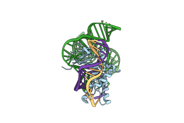
Isdra2 Tnpb In Complex With Rerna
Organism: Deinococcus radiodurans r1
Method: ELECTRON MICROSCOPY Release Date: 2023-04-12 Classification: RNA BINDING PROTEIN |
Organism: Deinococcus radiodurans r1
Method: ELECTRON MICROSCOPY
Release Date: 2023-04-12
 |
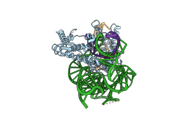
Isdra2 Tnpb In Complex With Rerna And Cognate Dna, Conformation 2 (Ruvc Domain Unresolved)
Organism: Deinococcus radiodurans r1, Synthetic construct
Method: ELECTRON MICROSCOPY Release Date: 2023-04-05 Classification: RNA BINDING PROTEIN |
Organism: Deinococcus radiodurans r1, Synthetic construct
Method: ELECTRON MICROSCOPY
Release Date: 2023-04-05
 |
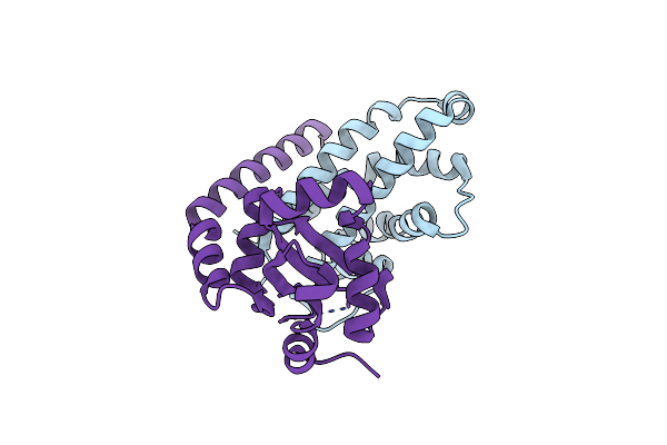
Isdra2 Tnpb In Complex With Rerna And Cognate Dna, Conformation 1 (Ruvc Domain Resolved)
Organism: Deinococcus radiodurans r1, Synthetic construct
Method: ELECTRON MICROSCOPY Release Date: 2023-04-05 Classification: RNA BINDING PROTEIN |
Organism: Deinococcus radiodurans r1, Synthetic construct
Method: ELECTRON MICROSCOPY
Release Date: 2023-04-05
 |
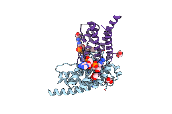
Crystal Structure Of Hepn(H107A-Y109F) Toxin In Complex With Mnt Antitoxin
Organism: Aphanizomenon flos-aquae 2012/km1/d3
Method: X-RAY DIFFRACTION Resolution:2.00 Å Release Date: 2020-12-30 Classification: TOXIN/ANTITOXIN |
Organism: Aphanizomenon flos-aquae 2012/km1/d3
Method: X-RAY DIFFRACTION
Release Date: 2020-12-30
 |
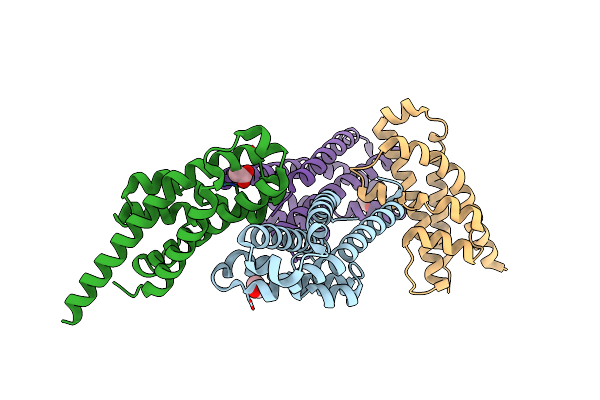
Crystal Structure Of Di-Ampylated Hepn(R102A) Toxin
Organism: Aphanizomenon flos-aquae 2012/km1/d3
Method: X-RAY DIFFRACTION Resolution:1.65 Å Release Date: 2020-12-30 Classification: TOXIN Ligands: AMP, EDO |
Organism: Aphanizomenon flos-aquae 2012/km1/d3
Method: X-RAY DIFFRACTION
Release Date: 2020-12-30
Ligands: AMP, EDO
 |
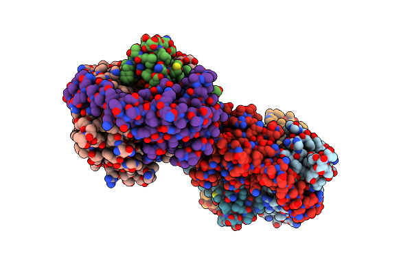
Crystal Structure Of Hepn(R102A) Toxin
Organism: Aphanizomenon flos-aquae 2012/km1/d3
Method: X-RAY DIFFRACTION Resolution:2.00 Å Release Date: 2020-12-30 Classification: TOXIN Ligands: ACT |
Organism: Aphanizomenon flos-aquae 2012/km1/d3
Method: X-RAY DIFFRACTION
Release Date: 2020-12-30
Ligands: ACT
 |
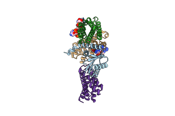
Crystal Structure Of Mono-Ampylated Hepn(R46E) Toxin In Complex With Mnt Antitoxin
Organism: Aphanizomenon flos-aquae 2012/km1/d3
Method: X-RAY DIFFRACTION Resolution:2.90 Å Release Date: 2020-12-30 Classification: TOXIN/ANTITOXIN Ligands: 2DA |
Organism: Aphanizomenon flos-aquae 2012/km1/d3
Method: X-RAY DIFFRACTION
Release Date: 2020-12-30
Ligands: 2DA
 |
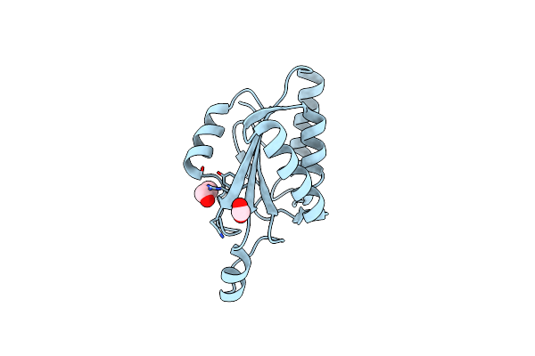
Rebuilt And Re-Refined Pdb Entry 5Yep: Tri-Ampylated Shewanella Oneidensis Hepn Toxin In Complex With Mnt Antitoxin
Organism: Shewanella oneidensis (strain mr-1)
Method: X-RAY DIFFRACTION Resolution:3.00 Å Release Date: 2020-12-30 Classification: TOXIN/ANTITOXIN Ligands: AMP Modified Residue(s): MSE |
Organism: Shewanella oneidensis (strain mr-1)
Method: X-RAY DIFFRACTION
Release Date: 2020-12-30
Ligands: AMP
 |
C-Terminal Domain Of Cgli Restriction Endonuclease H Subunit
Organism: Corynebacterium glutamicum
Method: X-RAY DIFFRACTION Resolution:2.40 Å Release Date: 2018-02-14 Classification: HYDROLASE Ligands: FMT, EDO Modified Residue(s): MSE |
Organism: Corynebacterium glutamicum
Method: X-RAY DIFFRACTION
Release Date: 2018-02-14
Ligands: FMT, EDO