
Search Count: 119
All
Selected
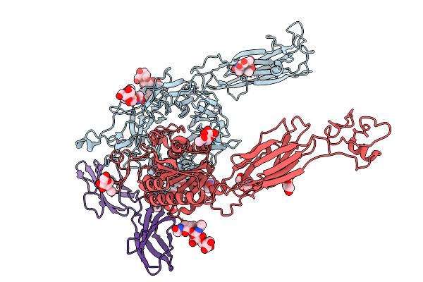 |
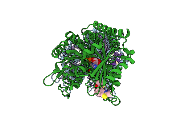
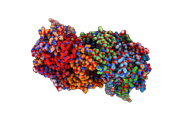
Cryo-Em Structure Of Human Integrin Alpha5Beta1 In Complex With Fibronectin (Fn 7-10)
Organism: Homo sapiens
Method: ELECTRON MICROSCOPY Resolution:2.61 Å Release Date: 2026-03-25 Classification: CELL ADHESION Ligands: MN, NAG |
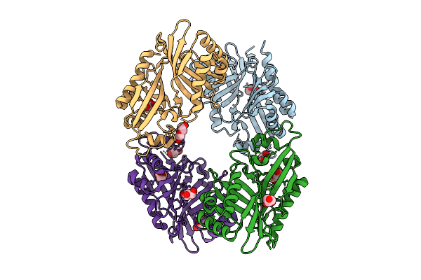 |
Organism: Streptomyces virginiae
Method: X-RAY DIFFRACTION Resolution:3.01 Å Release Date: 2025-12-10 Classification: HYDROLASE Ligands: GOL, PEG |
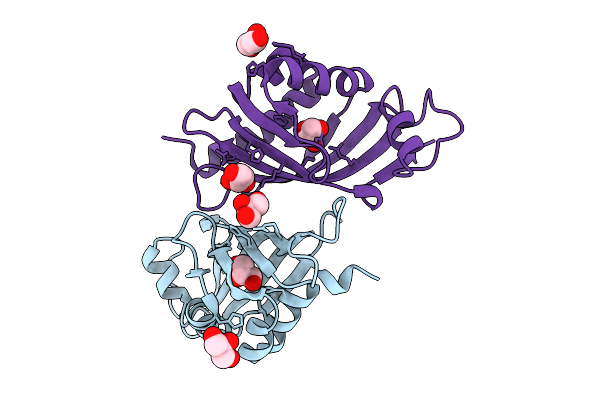 |
Organism: Streptomyces virginiae
Method: X-RAY DIFFRACTION Resolution:2.46 Å Release Date: 2025-12-10 Classification: HYDROLASE Ligands: GOL |
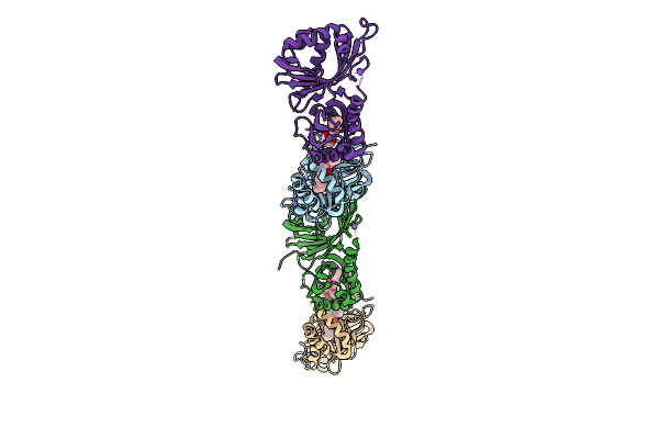 |
Organism: Streptomyces virginiae
Method: X-RAY DIFFRACTION Resolution:3.14 Å Release Date: 2025-12-10 Classification: HYDROLASE Ligands: A1L6P, NA |
 |
Organism: Mus musculus
Method: ELECTRON MICROSCOPY Resolution:3.19 Å Release Date: 2025-11-12 Classification: STRUCTURAL PROTEIN Ligands: MG, GDP, EPB, GTP |
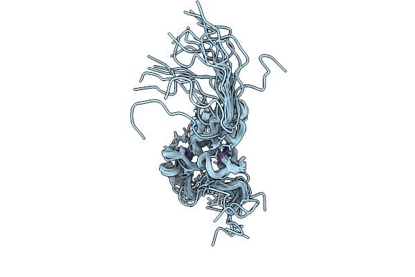 |
Organism: Measles morbillivirus
Method: SOLUTION NMR Release Date: 2025-09-03 Classification: VIRUS Ligands: ZN |
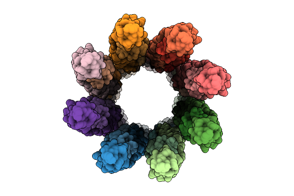 |
Organism: Caenorhabditis elegans
Method: ELECTRON MICROSCOPY Release Date: 2025-08-27 Classification: CELL CYCLE |
 |
Organism: Homo sapiens
Method: ELECTRON MICROSCOPY Resolution:3.84 Å Release Date: 2025-03-26 Classification: IMMUNE SYSTEM |
 |
Organism: Homo sapiens
Method: ELECTRON MICROSCOPY Resolution:3.07 Å Release Date: 2025-03-26 Classification: IMMUNE SYSTEM |
 |
Organism: Mus musculus, Oryctolagus cuniculus
Method: ELECTRON MICROSCOPY Release Date: 2025-02-12 Classification: CELL ADHESION Ligands: ADP, MG |
 |
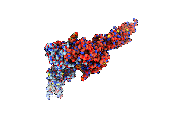
Organism: Severe acute respiratory syndrome coronavirus 2, Homo sapiens
Method: ELECTRON MICROSCOPY Release Date: 2024-07-31 Classification: VIRAL PROTEIN Ligands: NAG |
 |
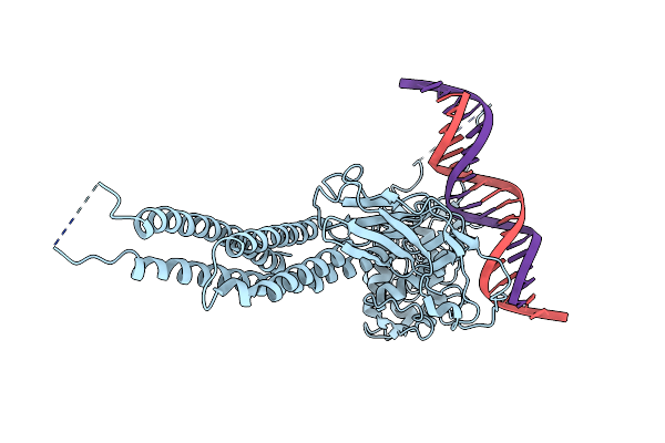
Homotetramic Antiparallel Coiled-Coil Of 23-Residues Laci C-Terminal Tetramerization Helix
Organism: Escherichia coli
Method: X-RAY DIFFRACTION Resolution:1.10 Å Release Date: 2023-08-30 Classification: DNA BINDING PROTEIN Ligands: 1PE |
 |
Organism: Escherichia coli
Method: X-RAY DIFFRACTION Resolution:1.54 Å Release Date: 2023-08-30 Classification: DE NOVO PROTEIN Ligands: GOL |
 |
Crystal Structure Of Bovine Heart Cytochrome C Oxidase, Apo Structure With Dmso
Organism: Bos taurus
Method: X-RAY DIFFRACTION Resolution:2.20 Å Release Date: 2022-12-21 Classification: OXIDOREDUCTASE Ligands: HEA, CU, MG, NA, PER, PGV, TGL, CUA, CDL, CHD, PEK, PSC, ZN, DMU |
 |
Crystal Structure Of Bovine Heart Cytochrome C Oxidase, The Structure Complexed With An Allosteric Inhibitor T113
Organism: Bos taurus
Method: X-RAY DIFFRACTION Resolution:2.20 Å Release Date: 2022-12-21 Classification: OXIDOREDUCTASE Ligands: HEA, CU, MG, NA, PER, TGL, J6X, CUA, CHD, PSC, PEK, PGV, CDL, DMU, ZN |
 |
Cryo-Em Structure Of Cytochrome Bo3 From Escherichia Coli, Apo Structure With Dmso
Organism: Escherichia coli
Method: ELECTRON MICROSCOPY Release Date: 2022-12-21 Classification: OXIDOREDUCTASE Ligands: HEO, HEM, CU, PEE, UNX |
 |
Cryo-Em Structure Of Cytochrome Bo3 From Escherichia Coli, The Structure Complexed With An Allosteric Inhibitor N4
Organism: Escherichia coli
Method: ELECTRON MICROSCOPY Release Date: 2022-12-21 Classification: OXIDOREDUCTASE Ligands: HEO, HEM, CU, PEE, JYR, UNX |
 |
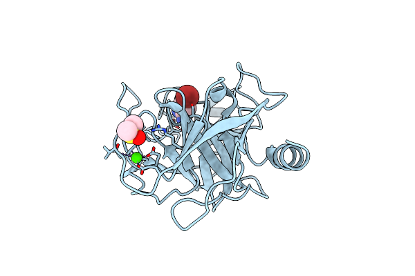
Crystal Structure Of Bovine Pancreatic Trypsin In Complex With Benzamidine At Room Temperature
Organism: Bos taurus
Method: X-RAY DIFFRACTION Resolution:1.77 Å Release Date: 2022-06-15 Classification: HYDROLASE Ligands: BEN, DMS, CA |
 |
Crystal Structure Of Bovine Pancreatic Trypsin In Complex With 4-Methoxybenzamidine At Room Temperature
Organism: Bos taurus
Method: X-RAY DIFFRACTION Resolution:1.52 Å Release Date: 2022-06-15 Classification: HYDROLASE Ligands: RKX, DMS, CA |
 |
Crystal Structure Of Bovine Pancreatic Trypsin In Complex With 4-Bromobenzamidine At Room Temperature
Organism: Bos taurus
Method: X-RAY DIFFRACTION Resolution:1.48 Å Release Date: 2022-06-15 Classification: HYDROLASE Ligands: F5R, DMS, CA |