Abstact
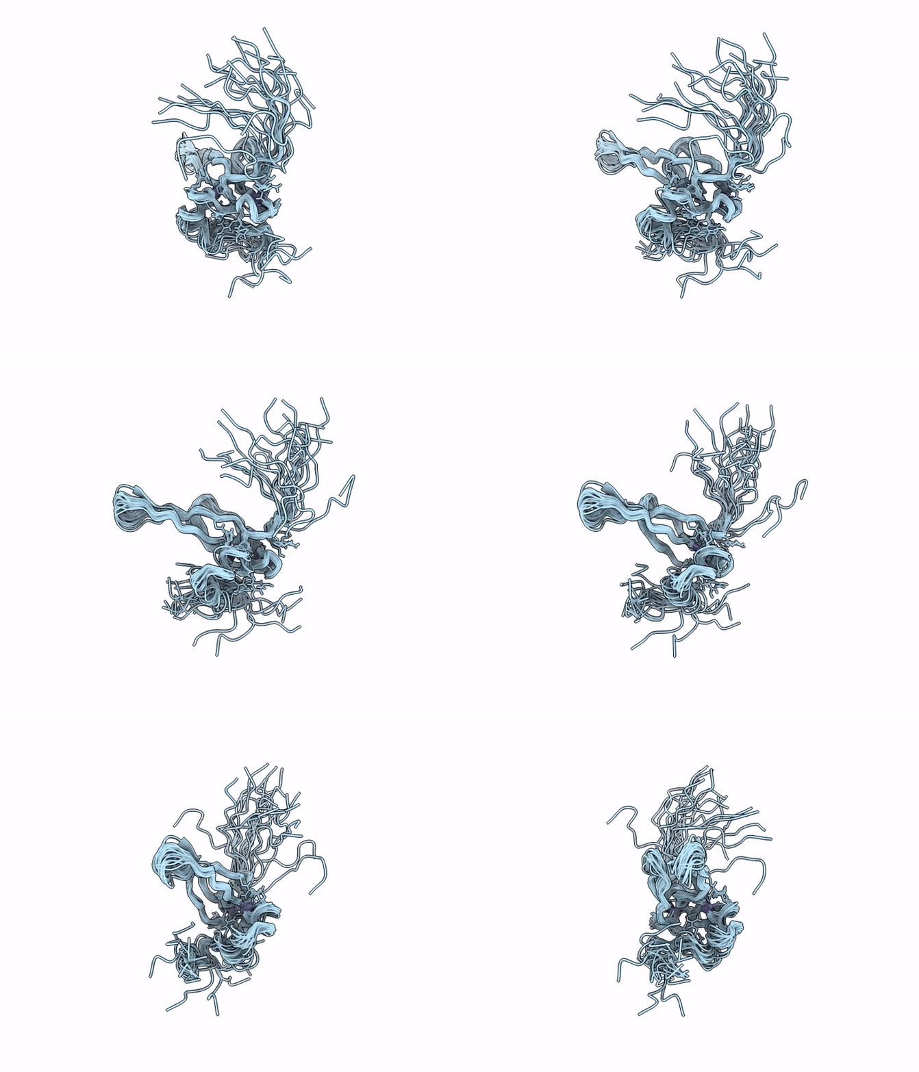
Viruses commonly evade the host antiviral interferon (IFN) response by targeting key components of the Janus kinase-signal transducer and activator of transcription (JAK-STAT) pathway, typically STAT1 and STAT2. Among the well-characterized viral IFN antagonists, measles virus (MeV), a member of the Morbillivirus genus, encodes a multifunctional V protein (MeV-V) that directly interacts with STAT proteins. The C-terminal domain (CTD) of MeV-V selectively binds to STAT2, disrupting the formation of the IFN-stimulated gene factor 3 (ISGF3) complex by inhibiting the STAT2-interferon regulatory factor 9 (IRF9) association. Here, we report a solution structure covering the MeV-V(CTD) in its unbound form, as determined by nuclear magnetic resonance spectroscopy. While the overall architecture, including a distinctive zinc-finger motif, conforms to previously predicted features, our analysis reveals unexpected features, including distinct proline cis conformers that may have functional relevance. Molecular mapping analysis, combined with relaxation measurements, identified key residues implicated in STAT2 recognition and revealed substantial conformational flexibility within the domain. These findings suggest that MeV-V(CTD) employs a shared binding surface for STAT2 binding as for melanoma differentiation-associated protein 5 (MDA5) interaction, underscoring its structural adaptability. As V proteins across Morbillivirus species engage diverse host pathways, including immune signaling, cell cycle regulation, and apoptosis, by targeting multiple proteins, we propose that the dynamic yet folded nature of the V(CTD) underlies its ability to serve as a versatile interaction module in host-pathogen interplay.IMPORTANCEThe measles virus V protein, encoded by the P gene, orchestrates the broad modulation of host responses, including immune evasion, by interacting with multiple host factors. With regard to structural studies of V(CTD), to date, only one protein from parainfluenza virus 5 has been crystallographically analyzed in complex with host targets. Despite the conserved nature of the V(CTD) among paramyxoviruses, structural information on the unbound state of this domain is lacking, and current insights largely rely on computational predictions based on the structure of the bound form. Our nuclear magnetic resonance work provides the first structure of the V(CTD) from paramyxoviruses in its free form. In accordance with our previously presented data, we further confirmed that the MeV-V binding site of STAT2 overlaps that of IRF9. The conformational flexibility observed within the folded CTD provides the structural basis for its ability to engage with multiple host targets with high specificity.