

Search Count: 76
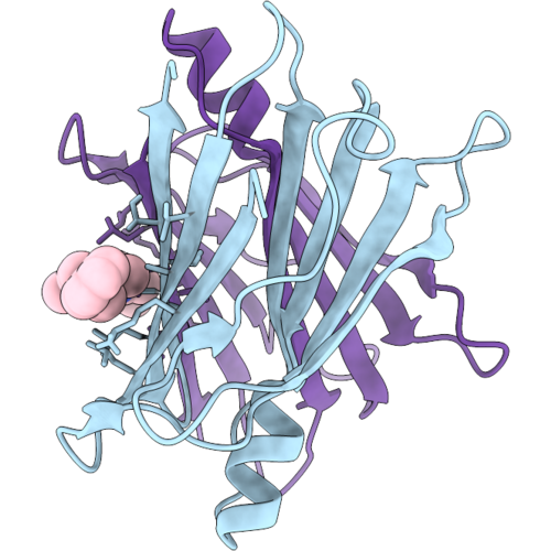 |
Discovery Of Cns Penetrant Transthyretin (Ttr) Stabilizers With Heterocyclic Linkers
Organism: Homo sapiens
Method: X-RAY DIFFRACTION Resolution:1.16 Å Release Date: 2026-10-07 Classification: TRANSPORT PROTEIN Ligands: A1DMZ |
Organism: Homo sapiens
Method: X-RAY DIFFRACTION
Release Date: 2026-10-07
Ligands: A1DMZ
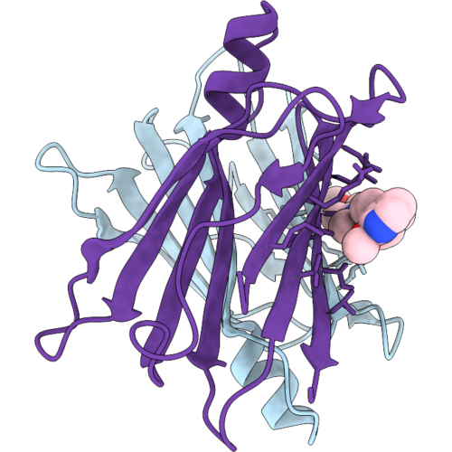 |
Discovery Of Cns Penetrant Transthyretin (Ttr) Stabilizers With Heterocyclic Linkers
Organism: Homo sapiens
Method: X-RAY DIFFRACTION Resolution:1.65 Å Release Date: 2026-10-07 Classification: TRANSPORT PROTEIN Ligands: A1DM0 |
Organism: Homo sapiens
Method: X-RAY DIFFRACTION
Release Date: 2026-10-07
Ligands: A1DM0
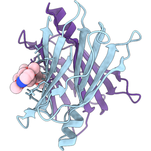 |
Discovery Of Neutral And Csf Penetrant Transthyretin (Ttr) Stabilizers With Heterocyclic Linkers
Organism: Homo sapiens
Method: X-RAY DIFFRACTION Resolution:1.28 Å Release Date: 2026-10-07 Classification: TRANSPORT PROTEIN Ligands: A1DM6 |
Organism: Homo sapiens
Method: X-RAY DIFFRACTION
Release Date: 2026-10-07
Ligands: A1DM6
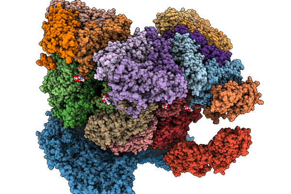 |
Rhesus Rotavirus (Consensus Structure At 4.7 Angstrom Resolution From Cryo-Et)
Organism: Simian rotavirus a strain rrv
Method: ELECTRON MICROSCOPY Release Date: 2026-03-04 Classification: VIRUS Ligands: NAG, CA, ZN, CL |
Organism: Simian rotavirus a strain rrv
Method: ELECTRON MICROSCOPY
Release Date: 2026-03-04
Ligands: NAG, CA, ZN, CL
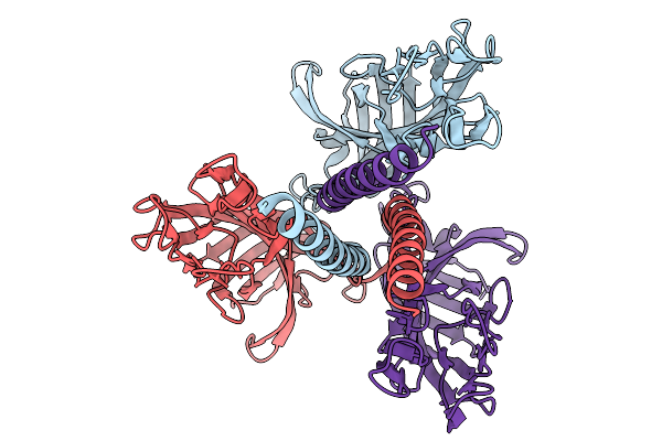 |
Membrane-Bound, Reversed Vp5* Trimer (Rotavirus Spike Protein)
Organism: Simian rotavirus a strain rrv
Method: ELECTRON MICROSCOPY Release Date: 2026-02-04 Classification: VIRAL PROTEIN |
Organism: Simian rotavirus a strain rrv
Method: ELECTRON MICROSCOPY
Release Date: 2026-02-04
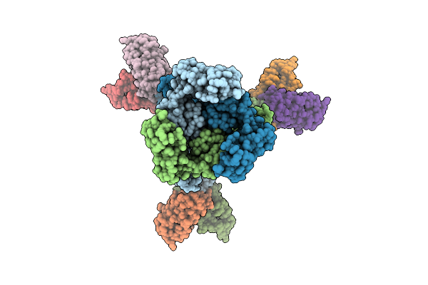 |
Structure Of A Murine Monoclonal Antibody Fab5 Targeting Epstein-Barr Virus Gb
Organism: Homo sapiens, Human gammaherpesvirus 4
Method: ELECTRON MICROSCOPY Release Date: 2025-10-15 Classification: VIRAL PROTEIN/IMMUNE SYSTEM Ligands: NAG |
Organism: Homo sapiens, Human gammaherpesvirus 4
Method: ELECTRON MICROSCOPY
Release Date: 2025-10-15
Ligands: NAG
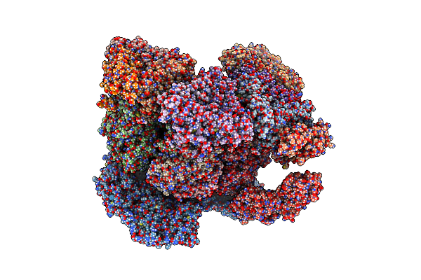 |
Rhesus Rotavirus (Consensus Structure At 2.36 Angstrom Resolution)
Organism: Simian rotavirus a strain rrv
Method: ELECTRON MICROSCOPY Release Date: 2025-04-30 Classification: VIRUS Ligands: NAG, CA, ZN, CL |
Organism: Simian rotavirus a strain rrv
Method: ELECTRON MICROSCOPY
Release Date: 2025-04-30
Ligands: NAG, CA, ZN, CL
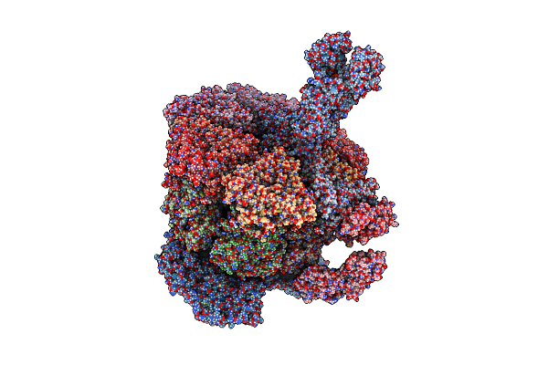 |
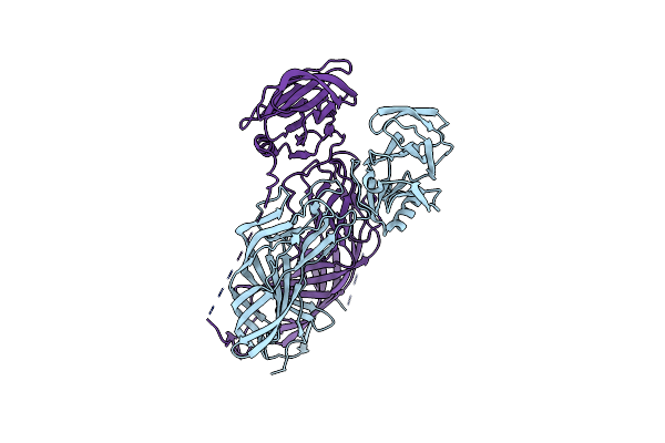
Rhesus Rotavirus (Upright Structure At 2.88 Angstrom Resolution)
Organism: Simian rotavirus a strain rrv
Method: ELECTRON MICROSCOPY Release Date: 2025-04-30 Classification: VIRAL PROTEIN Ligands: NAG, CA, ZN, CL |
Organism: Simian rotavirus a strain rrv
Method: ELECTRON MICROSCOPY
Release Date: 2025-04-30
Ligands: NAG, CA, ZN, CL
 |
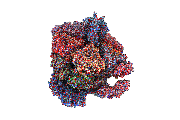
Rhesus Rotavirus (Vp5*/Vp8* Structure At 3.52 Angstrom Resolution)
Organism: Simian rotavirus a strain rrv
Method: ELECTRON MICROSCOPY Release Date: 2025-04-30 Classification: VIRAL PROTEIN |
Organism: Simian rotavirus a strain rrv
Method: ELECTRON MICROSCOPY
Release Date: 2025-04-30
 |
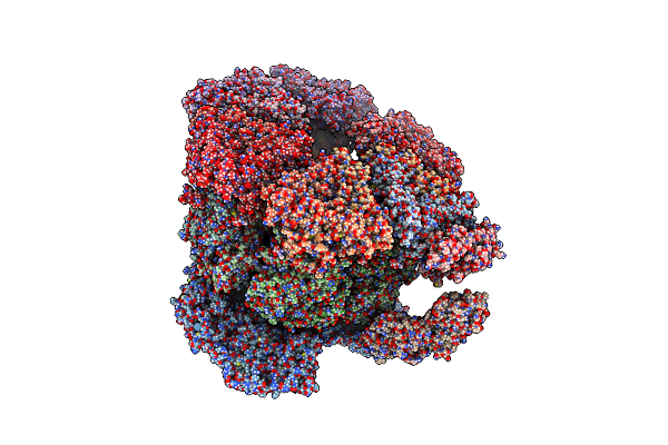
Rhesus Rotavirus (Reversed Structure At 2.72 Angstrom Resolution)
Organism: Simian rotavirus a strain rrv
Method: ELECTRON MICROSCOPY Release Date: 2025-04-30 Classification: VIRAL PROTEIN Ligands: NAG, CA, ZN, CL |
Organism: Simian rotavirus a strain rrv
Method: ELECTRON MICROSCOPY
Release Date: 2025-04-30
Ligands: NAG, CA, ZN, CL
 |
Rhesus Rotavirus (Empty Structure At 2.68 Angstrom Resolution)
Organism: Simian rotavirus a strain rrv
Method: ELECTRON MICROSCOPY Release Date: 2025-04-30 Classification: VIRAL PROTEIN Ligands: NAG, CA, ZN, CL |
Organism: Simian rotavirus a strain rrv
Method: ELECTRON MICROSCOPY
Release Date: 2025-04-30
Ligands: NAG, CA, ZN, CL
 |
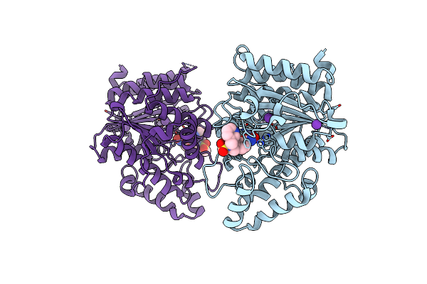
Rhesus Rotavirus (Vp1 Structure At 2.65 Angstrom Resolution)
Organism: Simian rotavirus a strain rrv
Method: ELECTRON MICROSCOPY Release Date: 2025-04-30 Classification: VIRAL PROTEIN |
Organism: Simian rotavirus a strain rrv
Method: ELECTRON MICROSCOPY
Release Date: 2025-04-30
 |
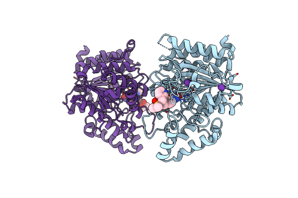
Crystal Structure Of Danio Rerio Histone Deacetylase 6 Catalytic Domain 2 Complexed With To-588
Organism: Danio rerio
Method: X-RAY DIFFRACTION Resolution:1.26 Å Release Date: 2024-12-04 Classification: METAL BINDING PROTEIN Ligands: A1BHY, ZN, K |
Organism: Danio rerio
Method: X-RAY DIFFRACTION
Release Date: 2024-12-04
Ligands: A1BHY, ZN, K
 |
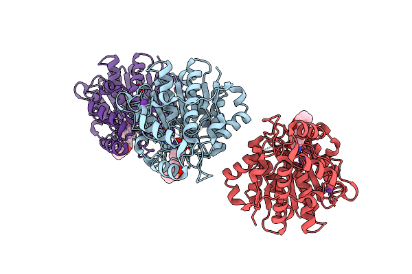
Crystal Structure Of Danio Rerio Histone Deacetylase 6 Catalytic Domain 2 Complexed With To-600
Organism: Danio rerio
Method: X-RAY DIFFRACTION Resolution:1.24 Å Release Date: 2024-12-04 Classification: METAL BINDING PROTEIN Ligands: A1BHX, ZN, K |
Organism: Danio rerio
Method: X-RAY DIFFRACTION
Release Date: 2024-12-04
Ligands: A1BHX, ZN, K
 |
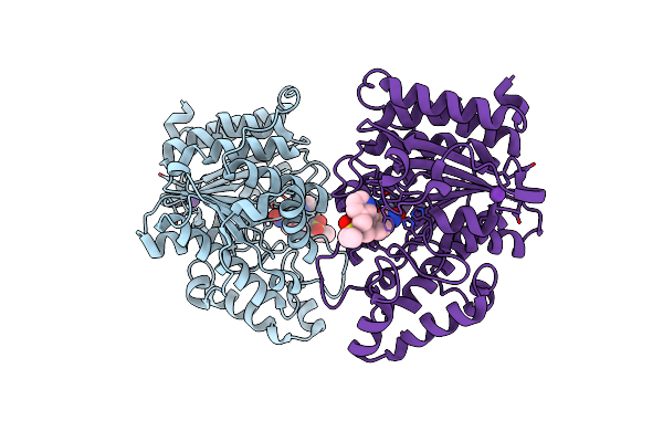
Crystal Structure Of Danio Rerio Histone Deacetylase 6 Catalytic Domain 2 Complexed With To-584
Organism: Danio rerio
Method: X-RAY DIFFRACTION Resolution:1.26 Å Release Date: 2024-12-04 Classification: METAL BINDING PROTEIN Ligands: ZN, A1BHW, K |
Organism: Danio rerio
Method: X-RAY DIFFRACTION
Release Date: 2024-12-04
Ligands: ZN, A1BHW, K
 |
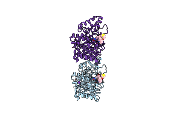
Crystal Structure Of Danio Rerio Histone Deacetylase 6 Catalytic Domain 2 Complexed With To-589
Organism: Danio rerio
Method: X-RAY DIFFRACTION Resolution:1.25 Å Release Date: 2024-12-04 Classification: METAL BINDING PROTEIN Ligands: A1BH8, ZN, K |
Organism: Danio rerio
Method: X-RAY DIFFRACTION
Release Date: 2024-12-04
Ligands: A1BH8, ZN, K
 |
Crystal Structure Of Danio Rerio Histone Deacetylase 6 Catalytic Domain 2 Complexed With To-599
Organism: Danio rerio
Method: X-RAY DIFFRACTION Resolution:1.27 Å Release Date: 2024-12-04 Classification: METAL BINDING PROTEIN Ligands: A1BH9, ZN, K |
Organism: Danio rerio
Method: X-RAY DIFFRACTION
Release Date: 2024-12-04
Ligands: A1BH9, ZN, K
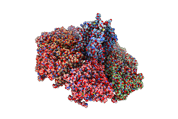 |
The Rotavirus Vp5*/Vp8* Conformational Transition Permeabilizes Membranes To Ca2+ (Class 5 Reconstruction)
Organism: Simian rotavirus a strain rrv
Method: ELECTRON MICROSCOPY Release Date: 2024-03-27 Classification: VIRAL PROTEIN Ligands: NAG, CA |
Organism: Simian rotavirus a strain rrv
Method: ELECTRON MICROSCOPY
Release Date: 2024-03-27
Ligands: NAG, CA
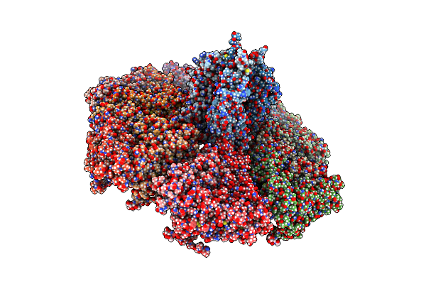 |
The Rotavirus Vp5*/Vp8* Conformational Transition Permeabilizes Membranes To Ca2+ (Class 6 Reconstruction)
Organism: Simian rotavirus a strain rrv
Method: ELECTRON MICROSCOPY Release Date: 2024-03-27 Classification: VIRAL PROTEIN Ligands: NAG, CA |
Organism: Simian rotavirus a strain rrv
Method: ELECTRON MICROSCOPY
Release Date: 2024-03-27
Ligands: NAG, CA
 |
Structure Of The Globular Isoform Of The Novel Conotoxin Pnid Derived From Conus Pennaceus
|
Organism: Conus pennaceus
Method: SOLUTION NMR
Release Date: 2023-01-25