

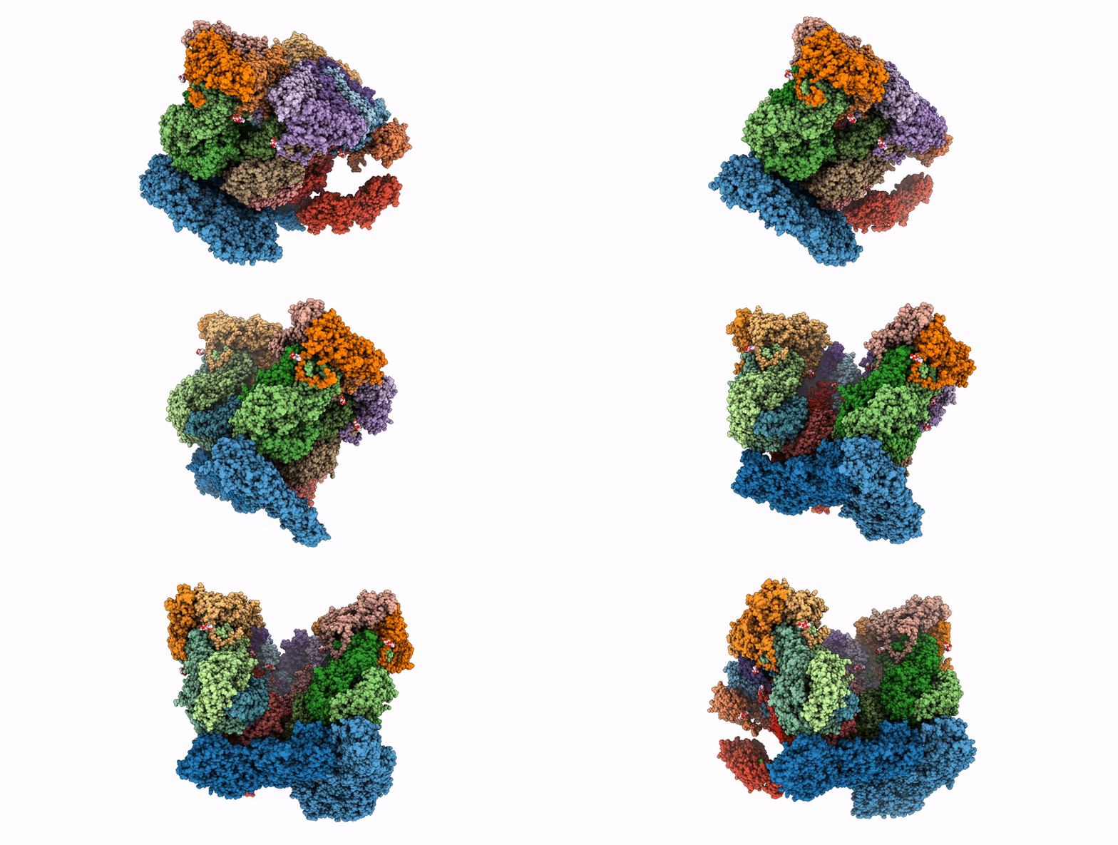
10IC image
Deposition Date 2026-01-21
Release Date 2026-03-04
Last Version Date 2026-03-04
Entry Detail
PDB ID:
10IC
Keywords:
Title:
Rhesus rotavirus (consensus structure at 4.7 Angstrom resolution from cryo-ET)
Biological Source:
Source Organism(s):
Expression System(s):
Method Details:
Experimental Method:
Resolution:
4.70 Å
Aggregation State:
CELL
Reconstruction Method:
SUBTOMOGRAM AVERAGING
Macromolecular Entities
Structures with similar UniProt ID
Protein Blast
Polymer Type:polypeptide(L)
Molecule:Outer capsid glycoprotein VP7
Chain IDs:A (auth: 0), B (auth: Y), C (auth: 1), D (auth: Z), T (auth: P), U (auth: Q), V (auth: R), W (auth: S), X (auth: T), Y (auth: U), Z (auth: V), AA (auth: W), BA (auth: X)
Chain Length:326
Number of Molecules:13
Biological Source:Simian rotavirus A strain RRV
Structures with similar UniProt ID
Protein Blast
Polymer Type:polypeptide(L)
Molecule:Inner capsid protein VP2
Chain IDs:E (auth: A), F (auth: B)
Chain Length:887
Number of Molecules:2
Biological Source:Simian rotavirus A strain RRV
Structures with similar UniProt ID
Protein Blast
Polymer Type:polypeptide(L)
Molecule:Intermediate capsid protein V
Chain IDs:G (auth: C), H (auth: D), I (auth: E), J (auth: F), K (auth: G), L (auth: H), M (auth: I), N (auth: J), O (auth: K), P (auth: L), Q (auth: M), R (auth: N), S (auth: O)
Chain Length:397
Number of Molecules:13
Biological Source:Simian rotavirus A strain RRV
Modified Residue
Compound ID Chain ID Parent Comp ID Details 2D Image
FME G MET modified residue
Primary Citation
Mechanism of membrane perforation in rotavirus cell entry.
Biorxiv ? ? ? (2026)
PMID: 41659395 DOI: 10.64898/2026.01.21.700916

Abstact

Infectious cell entry by non-enveloped viruses requires delivery of the viral genome -- in many cases enclosed within a large, subviral particle -- across the membrane of an intracellular compartment. Rotaviruses and other double-strand RNA (dsRNA) viruses introduce into their target cells an inner capsid particle, roughly 700 A in diameter, that does not uncoat further but instead extrudes capped viral mRNA by virtue of RNA-dependent RNA polymerase and capping activities within it. The delivery agent is an outer protein layer of the virion. We describe here use of cryogenic electron tomography (cryo-ET) to visualize the full course of rhesus rotavirus (RRV) entry, from cell attachment and inward budding of the virion to arrival of the subviral particle in the cytosol. The cryo-tomograms and subtomogram averaging of classified subparticles have enabled us to link high-resolution structures of the virion and its components with time series from live-cell fluorescence microscopy and thus to outline the molecular mechanism of each step in the entry process, including the hitherto elusive membrane perforation step needed for transfer of the subviral particle into the cytosol.

Legend

Protein

Chemical

Disease

Primary Citation of related structures
Feedback Form
Name
Email
Institute
Feedback