

Search Count: 37
 |
Characerization Of A Novel Format Scfvxvhh Single-Chain Biparatopic Antibody Against A Metal Binding Protein, Mtsa
Organism: Vicugna pacos, Streptococcus pyogenes
Method: X-RAY DIFFRACTION Resolution:3.66 Å Release Date: 2024-06-19 Classification: METAL TRANSPORT/IMMUNE SYSTEM |
Organism: Vicugna pacos, Streptococcus pyogenes
Method: X-RAY DIFFRACTION
Release Date: 2024-06-19
 |
Characerization Of A Novel Format Scfvxvhh Single-Chain Biparatopic Antibody Against A Metal Binding Protein, Mtsa
Organism: Streptococcus pyogenes
Method: X-RAY DIFFRACTION Resolution:1.37 Å Release Date: 2024-06-19 Classification: METAL TRANSPORT Ligands: ZN |
Organism: Streptococcus pyogenes
Method: X-RAY DIFFRACTION
Release Date: 2024-06-19
Ligands: ZN
 |
Characerization Of A Novel Format Scfvxvhh Single-Chain Biparatopic Antibody Against A Metal Binding Protein, Mtsa
Organism: Streptococcus pyogenes, Oryctolagus cuniculus
Method: X-RAY DIFFRACTION Resolution:2.80 Å Release Date: 2024-06-19 Classification: METAL TRNASPORT/IMMUNE SYSTEM Ligands: ZN |
Organism: Streptococcus pyogenes, Oryctolagus cuniculus
Method: X-RAY DIFFRACTION
Release Date: 2024-06-19
Ligands: ZN
 |
Characerization Of A Novel Format Scfvxvhh Single-Chain Biparatopic Antibody Against A Metal Binding Protein, Mtsa
Organism: Streptococcus pyogenes, Vicugna pacos
Method: X-RAY DIFFRACTION Resolution:1.65 Å Release Date: 2024-06-19 Classification: METAL TRANSPORT/IMMUNE SYSTEM Ligands: ZN |
Organism: Streptococcus pyogenes, Vicugna pacos
Method: X-RAY DIFFRACTION
Release Date: 2024-06-19
Ligands: ZN
 |
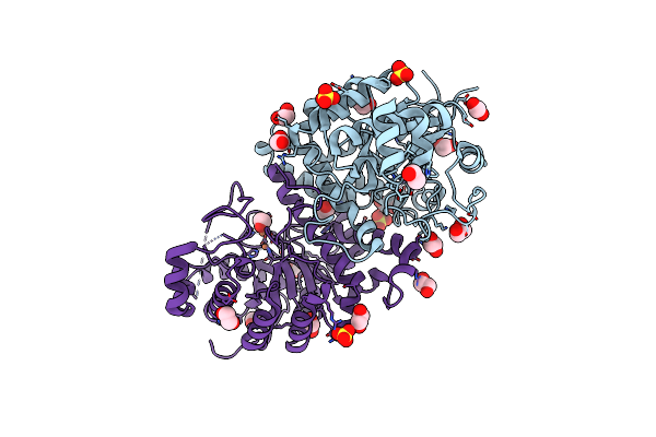
Structure Of The 2-Aminoisobutyric Acid Monooxygenase Hydroxylase
Organism: Rhodococcus wratislaviensis
Method: X-RAY DIFFRACTION Resolution:2.75 Å Release Date: 2021-01-06 Classification: OXIDOREDUCTASE Ligands: ZN, GOL, SO4, FE, CL Modified Residue(s): MSE |
Organism: Rhodococcus wratislaviensis
Method: X-RAY DIFFRACTION
Release Date: 2021-01-06
Ligands: ZN, GOL, SO4, FE, CL
 |
Structure Of The 2-Aminoisobutyric Acid Monooxygenase Hydroxylase
Organism: Rhodococcus wratislaviensis
Method: X-RAY DIFFRACTION Resolution:2.45 Å Release Date: 2021-01-06 Classification: OXIDOREDUCTASE Ligands: ZN, SO4, EDO, FE Modified Residue(s): MSE |
Organism: Rhodococcus wratislaviensis
Method: X-RAY DIFFRACTION
Release Date: 2021-01-06
Ligands: ZN, SO4, EDO, FE
 |
Crystal Structure Of Poz1 And Tpz1
Organism: Schizosaccharomyces pombe (strain 972 / atcc 24843) , Schizosaccharomyces pombe (strain 972 / atcc 24843)
Method: X-RAY DIFFRACTION Resolution:2.50 Å Release Date: 2017-12-20 Classification: DNA BINDING PROTEIN Ligands: SO4, ZN Modified Residue(s): MSE |
Organism: Schizosaccharomyces pombe (strain 972 / atcc 24843) , Schizosaccharomyces pombe (strain 972 / atcc 24843)
Method: X-RAY DIFFRACTION
Release Date: 2017-12-20
Ligands: SO4, ZN
 |
Crystal Structure Of Poz1, Tpz1 And Rap1
Organism: Schizosaccharomyces pombe (strain 972 / atcc 24843), Schizosaccharomyces pombe
Method: X-RAY DIFFRACTION Resolution:3.10 Å Release Date: 2017-12-20 Classification: DNA BINDING PROTEIN Ligands: ZN |
Organism: Schizosaccharomyces pombe (strain 972 / atcc 24843), Schizosaccharomyces pombe
Method: X-RAY DIFFRACTION
Release Date: 2017-12-20
Ligands: ZN
 |
Crystal Structure Of Hfa9 Egf Repeat With O-Glucose Trisaccharide
Organism: Homo sapiens
Method: X-RAY DIFFRACTION Resolution:2.20 Å Release Date: 2017-08-09 Classification: transferase, hydrolase Ligands: CA |
Organism: Homo sapiens
Method: X-RAY DIFFRACTION
Release Date: 2017-08-09
Ligands: CA
 |
Crystal Structure Of Drosophila Poglut1 (Rumi) Complexed With Its Glycoprotein Product (Glucosylated Egf Repeat) And Udp
Organism: Drosophila melanogaster, Homo sapiens
Method: X-RAY DIFFRACTION Resolution:2.50 Å Release Date: 2016-07-20 Classification: Transferase/Hydrolase Ligands: UDP, SO4, BGC |
Organism: Drosophila melanogaster, Homo sapiens
Method: X-RAY DIFFRACTION
Release Date: 2016-07-20
Ligands: UDP, SO4, BGC
 |
Crystal Structure Of Drosophila Poglut1 (Rumi) Complexed With Its Substrate Protein (Egf Repeat) And Udp
Organism: Drosophila melanogaster, Homo sapiens
Method: X-RAY DIFFRACTION Resolution:2.15 Å Release Date: 2016-07-20 Classification: TRANSFERASE/HYDROLASE Ligands: UDP, GOL, SO4 |
Organism: Drosophila melanogaster, Homo sapiens
Method: X-RAY DIFFRACTION
Release Date: 2016-07-20
Ligands: UDP, GOL, SO4
 |
Crystal Structure Of Drosophila Poglut1 (Rumi) Complexed With Its Substrate Protein (Egf Repeat)
Organism: Drosophila melanogaster, Homo sapiens
Method: X-RAY DIFFRACTION Resolution:1.90 Å Release Date: 2016-07-20 Classification: Transferase/Hydrolase Ligands: GOL, SO4 |
Organism: Drosophila melanogaster, Homo sapiens
Method: X-RAY DIFFRACTION
Release Date: 2016-07-20
Ligands: GOL, SO4
 |
Crystal Structure Of Drosophila Poglut1 (Rumi) Complexed With Udp
Organism: Drosophila melanogaster
Method: X-RAY DIFFRACTION Resolution:3.20 Å Release Date: 2016-07-20 Classification: TRANSFERASE Ligands: UDP, GOL |
Organism: Drosophila melanogaster
Method: X-RAY DIFFRACTION
Release Date: 2016-07-20
Ligands: UDP, GOL
 |
Crystal Structure Of Mouse Xyloside Xylosyltransferase 1, Apo Form
Organism: Mus musculus
Method: X-RAY DIFFRACTION Resolution:3.00 Å Release Date: 2015-11-04 Classification: TRANSFERASE Ligands: SO4 |
Organism: Mus musculus
Method: X-RAY DIFFRACTION
Release Date: 2015-11-04
Ligands: SO4
 |
Crystal Structure Of Mouse Xyloside Xylosyltransferase 1 Complexed With Manganese And Udp
Organism: Mus musculus
Method: X-RAY DIFFRACTION Resolution:3.03 Å Release Date: 2015-11-04 Classification: TRANSFERASE Ligands: MN, UDP, SO4 |
Organism: Mus musculus
Method: X-RAY DIFFRACTION
Release Date: 2015-11-04
Ligands: MN, UDP, SO4
 |
Crystal Structure Of Mouse Xyloside Xylosyltransferase 1 Complexed With Manganese
Organism: Mus musculus
Method: X-RAY DIFFRACTION Resolution:3.00 Å Release Date: 2015-09-30 Classification: TRANSFERASE Ligands: SO4, MN |
Organism: Mus musculus
Method: X-RAY DIFFRACTION
Release Date: 2015-09-30
Ligands: SO4, MN
 |
Crystal Structure Of Mouse Xyloside Xylosyltransferase 1 Complexed With Acceptor Ligand
Organism: Mus musculus, Homo sapiens
Method: X-RAY DIFFRACTION Resolution:2.37 Å Release Date: 2015-09-30 Classification: transferase/protein binding |
Organism: Mus musculus, Homo sapiens
Method: X-RAY DIFFRACTION
Release Date: 2015-09-30
 |
Crystal Structure Of Mouse Xyloside Xylosyltransferase 1 Complexed With Manganese,Acceptor Ligand And Udp-Glucose
Organism: Mus musculus, Homo sapiens
Method: X-RAY DIFFRACTION Resolution:1.62 Å Release Date: 2015-09-30 Classification: transferase/protein binding Ligands: MN, UPG, SO4 |
Organism: Mus musculus, Homo sapiens
Method: X-RAY DIFFRACTION
Release Date: 2015-09-30
Ligands: MN, UPG, SO4
 |
Crystal Structure Of Mouse Xyloside Xylosyltransferase 1 Complexed With Manganese, Acceptor Ligand And Udp
Organism: Mus musculus, Homo sapiens
Method: X-RAY DIFFRACTION Resolution:2.05 Å Release Date: 2015-09-30 Classification: transferase/protein binding Ligands: MN, UDP, SO4 |
Organism: Mus musculus, Homo sapiens
Method: X-RAY DIFFRACTION
Release Date: 2015-09-30
Ligands: MN, UDP, SO4
 |
Crystal Structure Of Mouse Xyloside Xylosyltransferase 1 Complexed With Manganese, Product Ligand And Udp (Product Complex I)
Organism: Mus musculus, Homo sapiens
Method: X-RAY DIFFRACTION Resolution:1.87 Å Release Date: 2015-09-30 Classification: transferase/protein binding Ligands: MN, UDP, SO4 |
Organism: Mus musculus, Homo sapiens
Method: X-RAY DIFFRACTION
Release Date: 2015-09-30
Ligands: MN, UDP, SO4