

Search Count: 28
 |
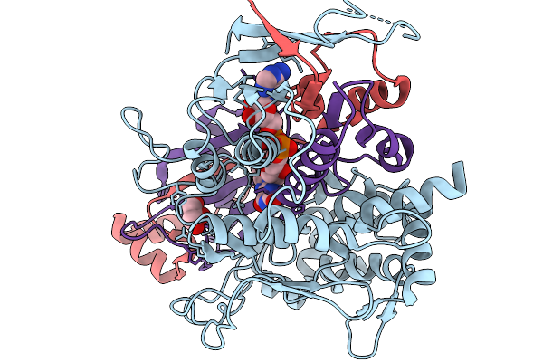
Structure Of L-Glutamate Oxidase In Complex With L-Glutamate
Organism: Streptomyces sp. x-119-6
Method: X-RAY DIFFRACTION Resolution:2.55 Å Release Date: 2026-01-07 Classification: OXIDOREDUCTASE Ligands: FAD, GLU |
Organism: Streptomyces sp. x-119-6
Method: X-RAY DIFFRACTION
Release Date: 2026-01-07
Ligands: FAD, GLU
 |
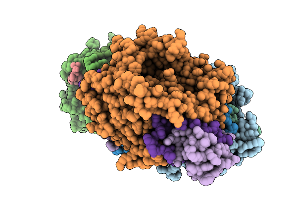
Structure Of L-Glutamate Oxidase E617Q Mutant
Organism: Streptomyces sp. x-119-6
Method: X-RAY DIFFRACTION Resolution:2.43 Å Release Date: 2026-01-07 Classification: OXIDOREDUCTASE Ligands: FAD, GOL |
Organism: Streptomyces sp. x-119-6
Method: X-RAY DIFFRACTION
Release Date: 2026-01-07
Ligands: FAD, GOL
 |
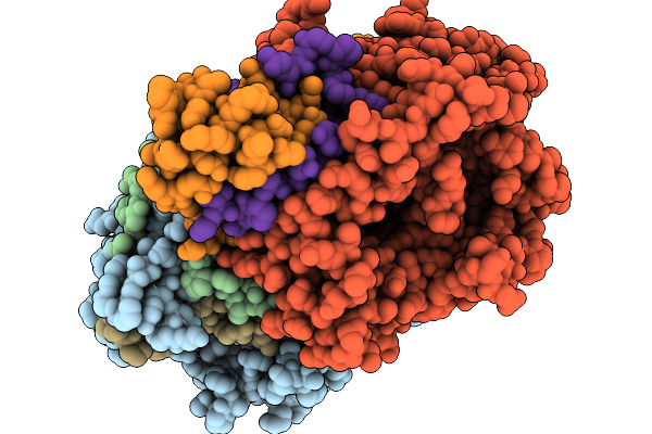
Structure Of L-Glutamate Oxidase E617Q Mutant In Complex With L-Glutamate
Organism: Streptomyces sp. x-119-6
Method: X-RAY DIFFRACTION Resolution:2.38 Å Release Date: 2026-01-07 Classification: OXIDOREDUCTASE Ligands: FAD, GLU |
Organism: Streptomyces sp. x-119-6
Method: X-RAY DIFFRACTION
Release Date: 2026-01-07
Ligands: FAD, GLU
 |
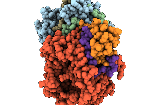
Structure Of L-Glutamate Oxidase E617F Mutant
Organism: Streptomyces sp. x-119-6
Method: X-RAY DIFFRACTION Resolution:3.27 Å Release Date: 2026-01-07 Classification: OXIDOREDUCTASE Ligands: FAD |
Organism: Streptomyces sp. x-119-6
Method: X-RAY DIFFRACTION
Release Date: 2026-01-07
Ligands: FAD
 |
Structure Of L-Glutamate Oxidase E617K Mutant
Organism: Streptomyces sp. x-119-6
Method: X-RAY DIFFRACTION Resolution:1.85 Å Release Date: 2026-01-07 Classification: OXIDOREDUCTASE Ligands: FAD |
Organism: Streptomyces sp. x-119-6
Method: X-RAY DIFFRACTION
Release Date: 2026-01-07
Ligands: FAD
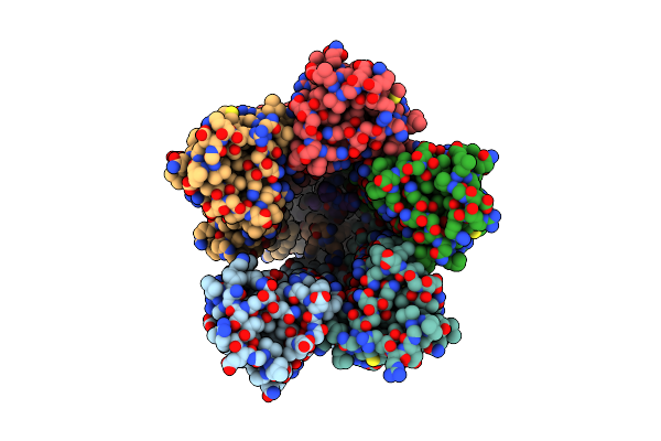 |
The Chimeric Flagellar Motor Complex Between Mota1B1 From Paenibacillus Sp. Tca20 And Motab From E.Coli, State 1
Organism: Paenibacillus sp. tca20, Escherichia coli
Method: ELECTRON MICROSCOPY Resolution:3.30 Å Release Date: 2025-04-09 Classification: MOTOR PROTEIN Ligands: AV0 |
Organism: Paenibacillus sp. tca20, Escherichia coli
Method: ELECTRON MICROSCOPY
Release Date: 2025-04-09
Ligands: AV0
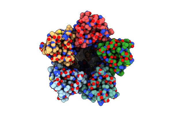 |
The Chimeric Flagellar Motor Complex Between Mota1B1 From Paenibacillus Sp. Tca20 And Motab From E.Coli, State 2
Organism: Paenibacillus sp. tca20, Escherichia coli
Method: ELECTRON MICROSCOPY Resolution:3.30 Å Release Date: 2025-04-09 Classification: MOTOR PROTEIN |
Organism: Paenibacillus sp. tca20, Escherichia coli
Method: ELECTRON MICROSCOPY
Release Date: 2025-04-09
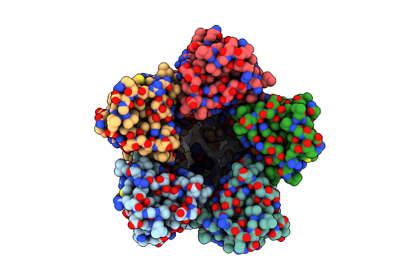 |
The Chimeric Flagellar Motor Complex Between Mota1B1 From Paenibacillus Sp. Tca20 And Motab From E.Coli, State 3
Organism: Paenibacillus sp. tca20
Method: ELECTRON MICROSCOPY Resolution:3.50 Å Release Date: 2025-04-09 Classification: MOTOR PROTEIN |
Organism: Paenibacillus sp. tca20
Method: ELECTRON MICROSCOPY
Release Date: 2025-04-09
 |
Structure Of The Periplasmic Domain Of Mots From Bacillus Subtilis
Organism: Bacillus subtilis
Method: X-RAY DIFFRACTION Resolution:1.89 Å Release Date: 2025-02-26 Classification: MOTOR PROTEIN Ligands: SO4 |
Organism: Bacillus subtilis
Method: X-RAY DIFFRACTION
Release Date: 2025-02-26
Ligands: SO4
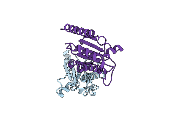 |
Structure Of The Periplasmic Domain Of Mots From Bacillus Subtilis In 300 Mm Nacl
Organism: Bacillus subtilis
Method: X-RAY DIFFRACTION Resolution:1.89 Å Release Date: 2025-02-26 Classification: MOTOR PROTEIN |
Organism: Bacillus subtilis
Method: X-RAY DIFFRACTION
Release Date: 2025-02-26
 |
Structure Of The Periplasmic Domain Of Mots From Bacillus Subtilis In 300 Mm Kcl
Organism: Bacillus subtilis
Method: X-RAY DIFFRACTION Resolution:1.90 Å Release Date: 2025-02-26 Classification: MOTOR PROTEIN |
Organism: Bacillus subtilis
Method: X-RAY DIFFRACTION
Release Date: 2025-02-26
 |
Bacterial Flagellar Sodium-Driven Stator Poma5Pomb2 With 100 Mm Nacl
Organism: Vibrio alginolyticus
Method: ELECTRON MICROSCOPY Release Date: 2025-01-01 Classification: MEMBRANE PROTEIN |
Organism: Vibrio alginolyticus
Method: ELECTRON MICROSCOPY
Release Date: 2025-01-01
 |
Bacterial Flagellar Sodium-Driven Stator Poma5Pomb2 With 100 Mm Kcl
Organism: Vibrio alginolyticus
Method: ELECTRON MICROSCOPY Release Date: 2025-01-01 Classification: MEMBRANE PROTEIN |
Organism: Vibrio alginolyticus
Method: ELECTRON MICROSCOPY
Release Date: 2025-01-01
 |
Bacterial Flagellar Sodium-Driven Stator Poma5Pomb2(D24N) With 100 Mm Nacl
Organism: Vibrio alginolyticus
Method: ELECTRON MICROSCOPY Release Date: 2025-01-01 Classification: MEMBRANE PROTEIN |
Organism: Vibrio alginolyticus
Method: ELECTRON MICROSCOPY
Release Date: 2025-01-01
 |
Bacterial Flagellar Sodium-Driven Stator Poma5Pomb2(D24N) With 100 Mm Kcl
Organism: Vibrio alginolyticus
Method: ELECTRON MICROSCOPY Release Date: 2025-01-01 Classification: MEMBRANE PROTEIN |
Organism: Vibrio alginolyticus
Method: ELECTRON MICROSCOPY
Release Date: 2025-01-01
 |
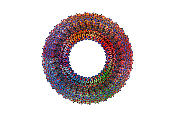
Bacterial Flagellar Sodium-Driven Stator Poma5Pomb2 With 100 Mm Nacl And 0.1 Mm Phenamil
Organism: Vibrio alginolyticus
Method: ELECTRON MICROSCOPY Release Date: 2025-01-01 Classification: MEMBRANE PROTEIN |
Organism: Vibrio alginolyticus
Method: ELECTRON MICROSCOPY
Release Date: 2025-01-01
 |
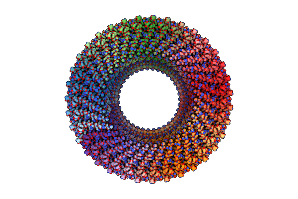
Structure Of The S-Ring Region Of The Vibrio Flagellar Ms-Ring Protein Flif With 34-Fold Symmetry Applied
Organism: Vibrio alginolyticus
Method: ELECTRON MICROSCOPY Release Date: 2024-09-04 Classification: MOTOR PROTEIN |
Organism: Vibrio alginolyticus
Method: ELECTRON MICROSCOPY
Release Date: 2024-09-04
 |
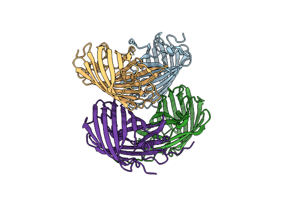
Structure Of The S-Ring Region Of The Vibrio Flagellar Ms-Ring Protein Flif With 35-Fold Symmetry Applied
Organism: Vibrio alginolyticus
Method: ELECTRON MICROSCOPY Release Date: 2024-09-04 Classification: MOTOR PROTEIN |
Organism: Vibrio alginolyticus
Method: ELECTRON MICROSCOPY
Release Date: 2024-09-04
 |
Structure Of Wild-Type Azami Green From Galaxea Fascicularis
Organism: Galaxea fascicularis
Method: X-RAY DIFFRACTION Resolution:1.62 Å Release Date: 2023-11-01 Classification: FLUORESCENT PROTEIN |
Organism: Galaxea fascicularis
Method: X-RAY DIFFRACTION
Release Date: 2023-11-01
 |
Structure Of Azami Red1.0, A Red Fluorescent Protein Engineered From Azami Green
Organism: Galaxea fascicularis
Method: X-RAY DIFFRACTION Resolution:1.84 Å Release Date: 2023-11-01 Classification: FLUORESCENT PROTEIN Ligands: CA |
Organism: Galaxea fascicularis
Method: X-RAY DIFFRACTION
Release Date: 2023-11-01
Ligands: CA