

Search Count: 20
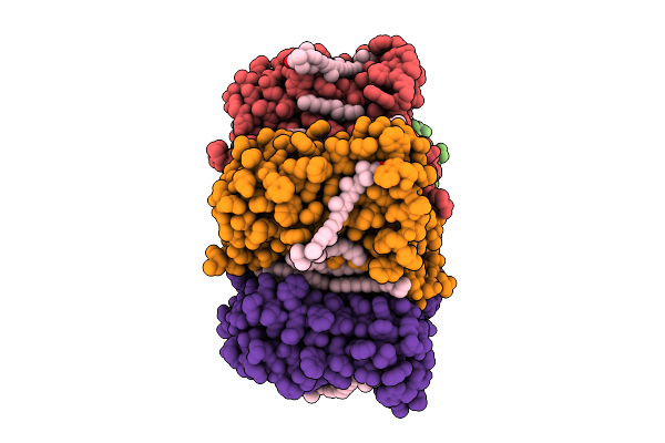 |
Cryo-Em Structure Of The Zeaxanthin-Bound Light-Driven Proton Pumping Rhodopsin, Nm-R1
Organism: Nonlabens marinus s1-08
Method: ELECTRON MICROSCOPY Resolution:2.46 Å Release Date: 2025-07-30 Classification: MEMBRANE PROTEIN Ligands: RET, R16, D12, K3I |
Organism: Nonlabens marinus s1-08
Method: ELECTRON MICROSCOPY
Release Date: 2025-07-30
Ligands: RET, R16, D12, K3I
 |
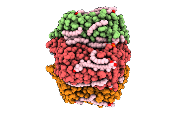
Cryo-Em Structure Of The Myxol-Bound Light-Driven Proton Pumping Rhodopsin, Nm-R1
Organism: Nonlabens marinus s1-08
Method: ELECTRON MICROSCOPY Resolution:2.27 Å Release Date: 2025-07-30 Classification: MEMBRANE PROTEIN Ligands: RET, R16, D12, A1L4O |
Organism: Nonlabens marinus s1-08
Method: ELECTRON MICROSCOPY
Release Date: 2025-07-30
Ligands: RET, R16, D12, A1L4O
 |
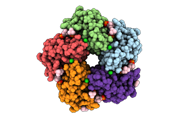
Cryo-Em Structure Of The Myxol-Bound Light-Driven Chloride Ion-Pumping Rhodopsin, Nm-R3
Organism: Nonlabens marinus s1-08
Method: ELECTRON MICROSCOPY Resolution:2.48 Å Release Date: 2025-07-30 Classification: MEMBRANE PROTEIN Ligands: RET, A1L4O, CL, PC1, PLC, R16, 8K6, D12, C14, D10 |
Organism: Nonlabens marinus s1-08
Method: ELECTRON MICROSCOPY
Release Date: 2025-07-30
Ligands: RET, A1L4O, CL, PC1, PLC, R16, 8K6, D12, C14, D10
 |
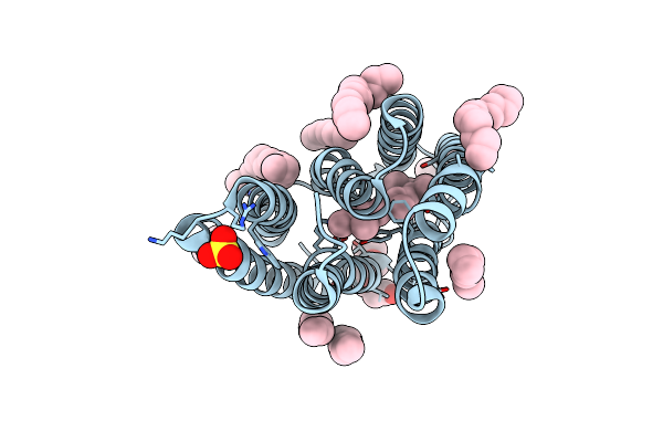
Cryo-Em Structure Of The Light-Driven Chloride Ion-Pumping Rhodopsin, Nm-R3
Organism: Nonlabens marinus s1-08
Method: ELECTRON MICROSCOPY Resolution:2.50 Å Release Date: 2025-07-30 Classification: MEMBRANE PROTEIN Ligands: RET, CL, PC1, PLC, D12, R16, 8K6, C14 |
Organism: Nonlabens marinus s1-08
Method: ELECTRON MICROSCOPY
Release Date: 2025-07-30
Ligands: RET, CL, PC1, PLC, D12, R16, 8K6, C14
 |
The Crystal Structure Of Cyanorhodopsin-Ii (Cyr-Ii) P7104R From Nodosilinea Nodulosa Pcc 7104
Organism: Nodosilinea nodulosa pcc 7104
Method: X-RAY DIFFRACTION Resolution:2.07 Å Release Date: 2023-10-25 Classification: MEMBRANE PROTEIN Ligands: RET, PG4, HEX, OCT, C14, R16, SO4, CL |
Organism: Nodosilinea nodulosa pcc 7104
Method: X-RAY DIFFRACTION
Release Date: 2023-10-25
Ligands: RET, PG4, HEX, OCT, C14, R16, SO4, CL
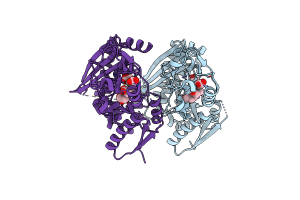 |
Crystal Structure Of The Holo Form Of Human Hydroxymethylbilane Synthase
Organism: Homo sapiens
Method: X-RAY DIFFRACTION Resolution:1.84 Å Release Date: 2021-03-17 Classification: TRANSFERASE Ligands: DPM |
Organism: Homo sapiens
Method: X-RAY DIFFRACTION
Release Date: 2021-03-17
Ligands: DPM
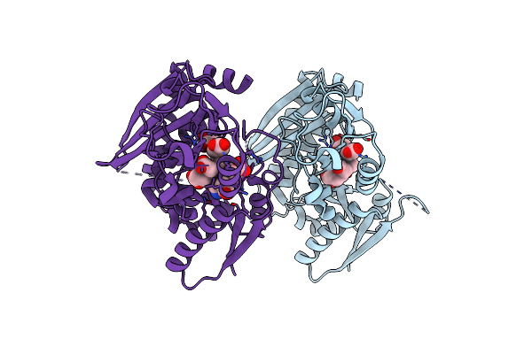 |
Crystal Structure Of The 2-Iodoporphobilinogen-Bound Holo Form Of Human Hydroxymethylbilane Synthase
Organism: Homo sapiens
Method: X-RAY DIFFRACTION Resolution:2.40 Å Release Date: 2021-03-17 Classification: TRANSFERASE Ligands: DPM, FWL |
Organism: Homo sapiens
Method: X-RAY DIFFRACTION
Release Date: 2021-03-17
Ligands: DPM, FWL
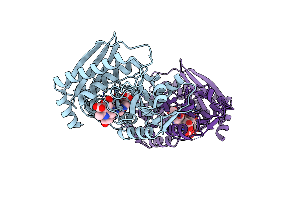 |
Crystal Structure Of The Es2 Intermediate Form Of Human Hydroxymethylbilane Synthase
Organism: Homo sapiens
Method: X-RAY DIFFRACTION Resolution:1.79 Å Release Date: 2021-03-17 Classification: TRANSFERASE Ligands: 7J8 |
Organism: Homo sapiens
Method: X-RAY DIFFRACTION
Release Date: 2021-03-17
Ligands: 7J8
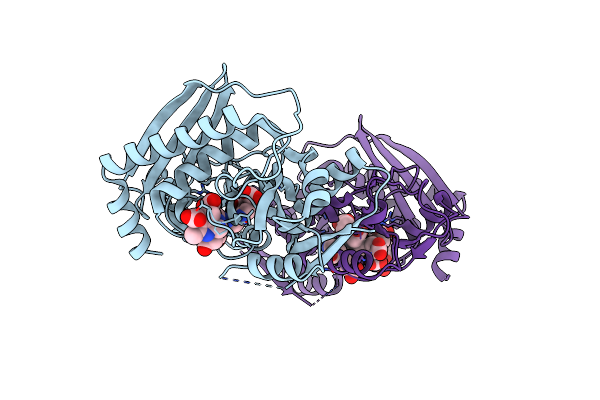 |
Crystal Structure Of The 2-Iodoporphobilinogen-Bound Es2 Intermediate Form Of Human Hydroxymethylbilane Synthase
Organism: Homo sapiens
Method: X-RAY DIFFRACTION Resolution:2.31 Å Release Date: 2021-03-17 Classification: TRANSFERASE Ligands: 7J8, FWL |
Organism: Homo sapiens
Method: X-RAY DIFFRACTION
Release Date: 2021-03-17
Ligands: 7J8, FWL
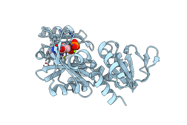 |
Crystal Structure Of Rimk Complexed With Rpsf
Organism: Escherichia coli k-12
Method: X-RAY DIFFRACTION Resolution:3.15 Å Release Date: 2019-03-27 Classification: LIGASE Ligands: ADP, MG |
Organism: Escherichia coli k-12
Method: X-RAY DIFFRACTION
Release Date: 2019-03-27
Ligands: ADP, MG
 |
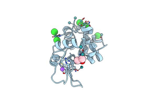
Crystal Structure Of Protease Anisep From Arthrobacter Nicotinovorans
Organism: Arthrobacter nicotinovorans
Method: X-RAY DIFFRACTION Resolution:1.70 Å Release Date: 2015-08-26 Classification: HYDROLASE |
Organism: Arthrobacter nicotinovorans
Method: X-RAY DIFFRACTION
Release Date: 2015-08-26
 |
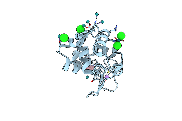
Crystal Structure Of Cross-Linked Tetragonal Hen Egg White Lysozyme Soaked With 5Mm [Ru(Co)3Cl2]2
Organism: Gallus gallus
Method: X-RAY DIFFRACTION Resolution:1.55 Å Release Date: 2014-12-31 Classification: HYDROLASE Ligands: DMF, CL, NA, RU1, RU |
Organism: Gallus gallus
Method: X-RAY DIFFRACTION
Release Date: 2014-12-31
Ligands: DMF, CL, NA, RU1, RU
 |
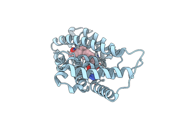
Crystal Structure Of Cross-Linked Tetragonal Hen Egg White Lysozyme Soaked With 5Mm [Ru(Co)3Cl2]2 Followed By The Reaction In Deoxy-Myoglobin Solution
Organism: Gallus gallus
Method: X-RAY DIFFRACTION Resolution:1.50 Å Release Date: 2014-12-31 Classification: HYDROLASE Ligands: DMF, CL, NA, RU1, RU |
Organism: Gallus gallus
Method: X-RAY DIFFRACTION
Release Date: 2014-12-31
Ligands: DMF, CL, NA, RU1, RU
 |
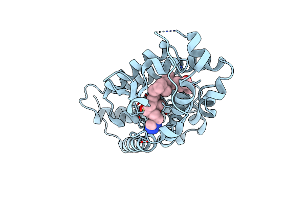
Crystal Structure Of The Human Vitamin D Receptor Ligand Binding Domain Complexed With 1Alpha,25-Dihydroxy-2Alpha-[2-(2H-Tetrazol-2-Yl)Ethyl]Vitamin D3
Organism: Homo sapiens
Method: X-RAY DIFFRACTION Resolution:2.49 Å Release Date: 2014-01-22 Classification: TRANSCRIPTION Ligands: TEY |
Organism: Homo sapiens
Method: X-RAY DIFFRACTION
Release Date: 2014-01-22
Ligands: TEY
 |
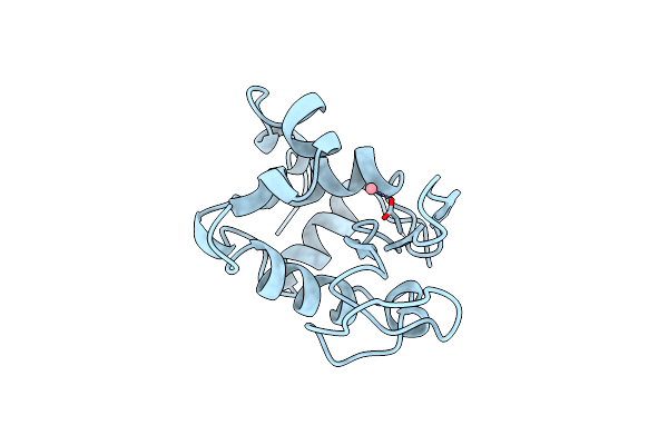
Crystal Structure Of The Human Vitamin D Receptor Ligand Binding Domain Complexed With 1Alpha,25-Dihydroxy-2Alpha-[2-(1H-Tetrazole-1-Yl)Ethyl]Vitamin D3
Organism: Homo sapiens
Method: X-RAY DIFFRACTION Resolution:2.84 Å Release Date: 2014-01-22 Classification: TRANSCRIPTION Ligands: TFY |
Organism: Homo sapiens
Method: X-RAY DIFFRACTION
Release Date: 2014-01-22
Ligands: TFY
 |
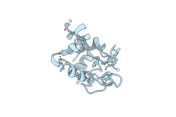
Crystal Structure Of Co/O-Hewl
Organism: Gallus gallus
Method: X-RAY DIFFRACTION Resolution:1.62 Å Release Date: 2012-05-09 Classification: HYDROLASE Ligands: CO, CL |
Organism: Gallus gallus
Method: X-RAY DIFFRACTION
Release Date: 2012-05-09
Ligands: CO, CL
 |
Crystal Structure Of Pt/O-Hewl
Organism: Gallus gallus
Method: X-RAY DIFFRACTION Resolution:2.34 Å Release Date: 2012-05-09 Classification: HYDROLASE Ligands: PT |
Organism: Gallus gallus
Method: X-RAY DIFFRACTION
Release Date: 2012-05-09
Ligands: PT
 |
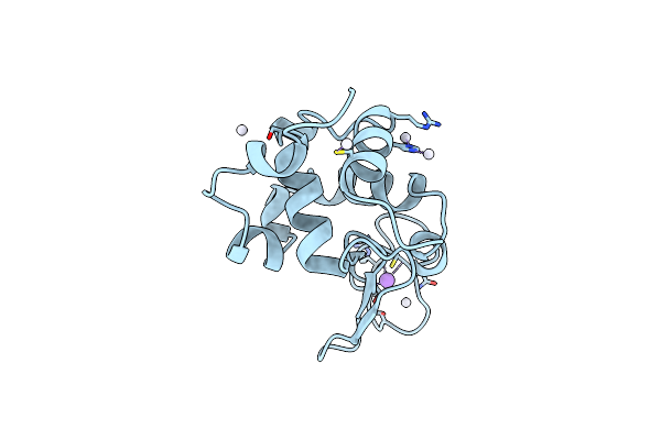
Crystal Structure Of Co/T-Hewl
Organism: Gallus gallus
Method: X-RAY DIFFRACTION Resolution:1.50 Å Release Date: 2012-05-09 Classification: HYDROLASE Ligands: CO, NA, CL, GOL |
Organism: Gallus gallus
Method: X-RAY DIFFRACTION
Release Date: 2012-05-09
Ligands: CO, NA, CL, GOL
 |
Crystal Structure Of Pt/T-Hewl
Organism: Gallus gallus
Method: X-RAY DIFFRACTION Resolution:2.10 Å Release Date: 2012-05-09 Classification: HYDROLASE Ligands: PT, NA |
Organism: Gallus gallus
Method: X-RAY DIFFRACTION
Release Date: 2012-05-09
Ligands: PT, NA
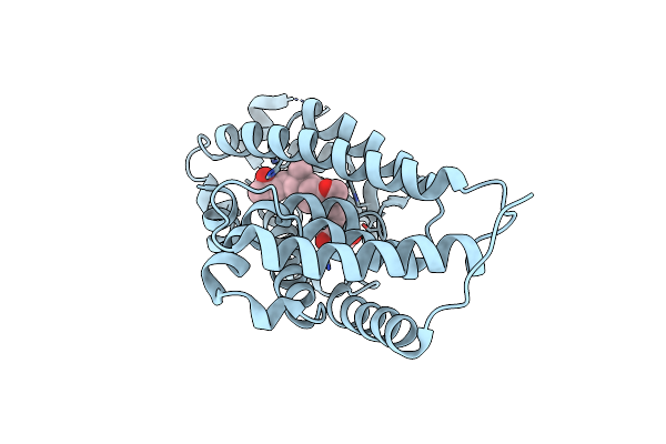 |
Crystal Structure Of The Human Vitamin D Receptor Ligand Binding Domain Complexed With 15Alpha-Methoxy-1Alpha,25-Dihydroxyvitamin D3
Organism: Homo sapiens
Method: X-RAY DIFFRACTION Resolution:2.60 Å Release Date: 2011-10-05 Classification: TRANSCRIPTION Ligands: EIM |
Organism: Homo sapiens
Method: X-RAY DIFFRACTION
Release Date: 2011-10-05
Ligands: EIM