
Search Count: 194
 |
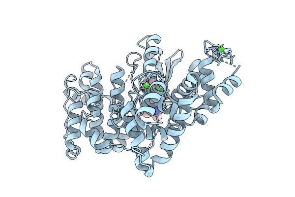
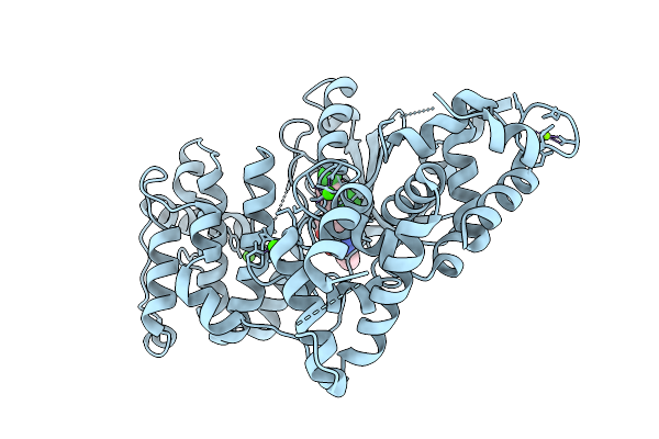
Akt1 Mutant In Complex With Miransertib (Arq 092)
Organism: Homo sapiens
Method: X-RAY DIFFRACTION Resolution:2.50 Å Release Date: 2026-07-01 Classification: TRANSFERASE Ligands: 6S1, EDO, GOL |
 |
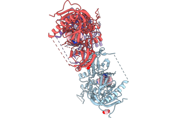
Sars-Cov-2 Rna-Dependent Rna Polymerase In Complex With 4'-Fla Nucleotide Analogue
Organism: Severe acute respiratory syndrome coronavirus 2
Method: ELECTRON MICROSCOPY Release Date: 2026-06-24 Classification: VIRAL PROTEIN/DNA Ligands: ZN, POP, A1DCZ, MG |
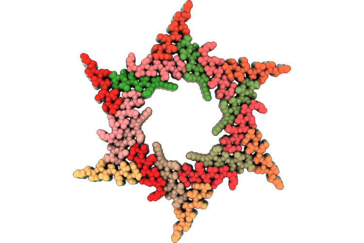 |
Hexagonally Ordered Nanofibrils Easily Yield By Dual Amphiphilic Self-Assembling Peptide
Organism: Other sequences
Method: ELECTRON MICROSCOPY Release Date: 2026-05-06 Classification: PROTEIN FIBRIL |
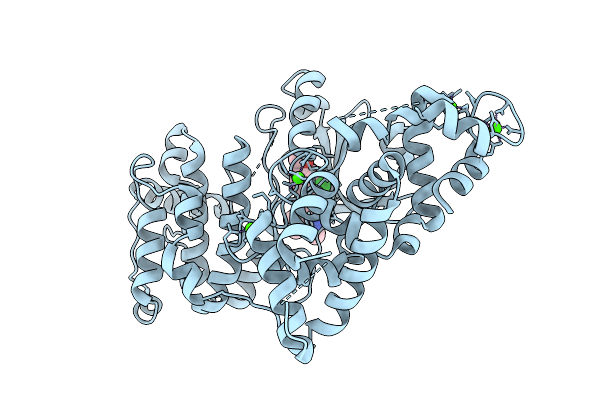 |
Crystal Structure Of Calcium-Dependent Protein Kinase 1 (Cdpk1) From Cryptosporidium Parvum In Complex With Inhibitor Win-1-158.
Organism: Cryptosporidium parvum iowa ii
Method: X-RAY DIFFRACTION Resolution:2.21 Å Release Date: 2026-04-01 Classification: TRANSFERASE Ligands: A1B1F, CA |
 |
Crystal Structure Of Calcium-Dependent Protein Kinase 1 (Cdpk1) From Cryptosporidium Parvum In Complex With Inhibitor Win-1-159.
Organism: Cryptosporidium parvum iowa ii
Method: X-RAY DIFFRACTION Resolution:2.30 Å Release Date: 2026-04-01 Classification: TRANSFERASE Ligands: A1B1U, CA |
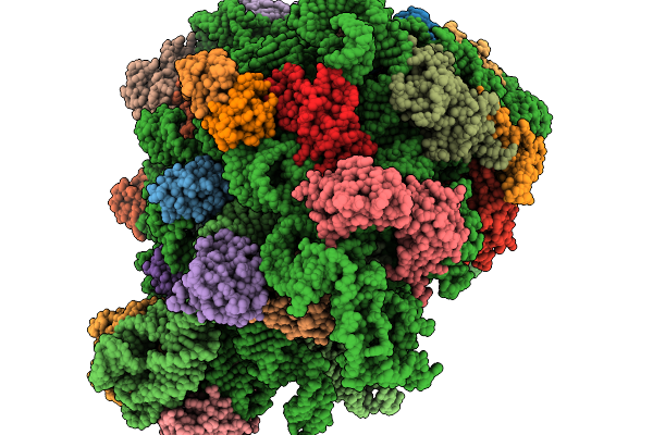 |
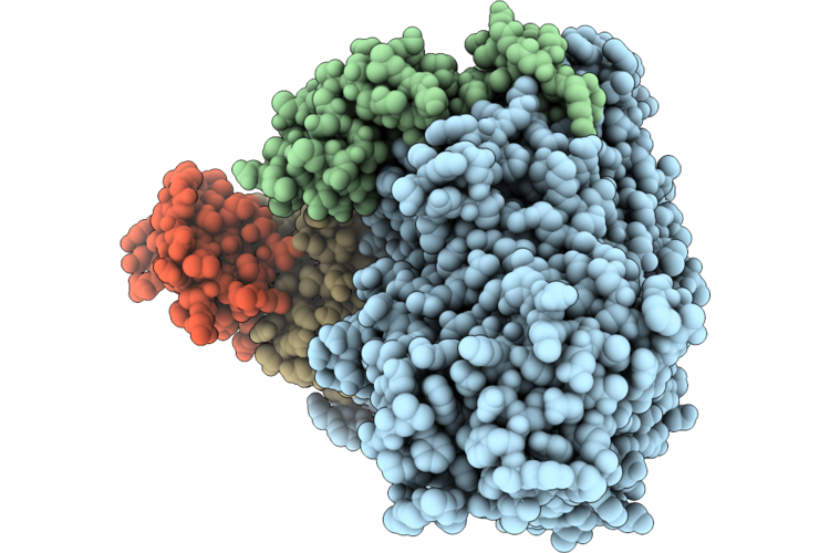
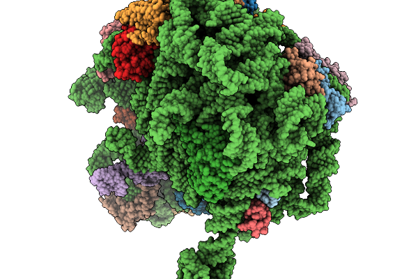
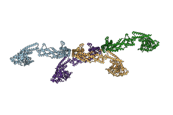
Structure Of E.Coli Ribosome With Filamin Mutant Y719E Nascent Chain At Linker Length Of 47 Amino Acids, With Trna
Organism: Dictyostelium discoideum, Escherichia coli
Method: ELECTRON MICROSCOPY Release Date: 2026-02-04 Classification: RIBOSOME |
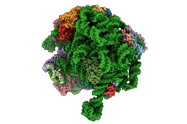 |
Structure Of E.Coli Ribosome With Nascent Chain At Linker Length Of 31 Amino Acids, With Mrna, P-Site And A-Site Trnas
Organism: Dictyostelium discoideum, Escherichia coli
Method: ELECTRON MICROSCOPY Release Date: 2026-01-28 Classification: RIBOSOME |
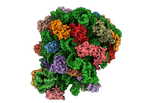 |
Structure Of Wt E.Coli Ribosome With Complexed Filament Nascent Chain At Length 34, With Mrna, P-Site And A-Site Trnas, And Mrna
Organism: Dictyostelium discoideum, Escherichia coli
Method: ELECTRON MICROSCOPY Release Date: 2026-01-21 Classification: RIBOSOME |
 |
Structure Of Wt E.Coli Ribosome With Complexed Filament Nascent Chain At Length 47, With P-Site Trna
Organism: Dictyostelium discoideum, Escherichia coli
Method: ELECTRON MICROSCOPY Release Date: 2026-01-14 Classification: RIBOSOME |
 |
Structure Of Wt E.Coli Ribosome With Complexed Filament Nascent Chain At Length 31, With P-Site Trnas
Organism: Dictyostelium discoideum, Escherichia coli
Method: ELECTRON MICROSCOPY Release Date: 2026-01-14 Classification: RIBOSOME |
 |
Crystal Structure Of Calcium-Dependent Protein Kinase 1 (Cdpk1) From Cryptosporidium Parvum In Complex With Inhibitor Win-3-115
Organism: Cryptosporidium parvum iowa ii
Method: X-RAY DIFFRACTION Resolution:2.94 Å Release Date: 2025-12-17 Classification: TRANSFERASE Ligands: CA, A1BLB, MG, CL |
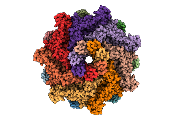 |
Aerolysin E254A/E258A In Styrene-Maleic Acid Lipid Particles
Organism: Aeromonas hydrophila
Method: ELECTRON MICROSCOPY Resolution:2.30 Å Release Date: 2025-11-26 Classification: TOXIN |
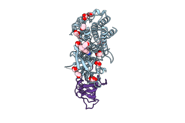 |
Cocrystal Structure Of Zilurgisertib Bound To The Alk2-Fkbp12 Complex
Organism: Homo sapiens
Method: X-RAY DIFFRACTION Resolution:1.75 Å Release Date: 2025-11-12 Classification: TRANSFERASE Ligands: A1JFB, EDO |
 |
Cryoem Structure Of Human S-Opa1 Assembled On Lipid Membrane Containing Brominated Cardiolipin In Membrane-Adjacent State
Organism: Homo sapiens
Method: ELECTRON MICROSCOPY Release Date: 2025-10-08 Classification: MEMBRANE PROTEIN |
 |
Cryoem Structure Of Human S-Opa1 Assembled On Lipid Membrane Containing Brominated Cardiolipin In Membrane-Adjacent State
Organism: Homo sapiens
Method: ELECTRON MICROSCOPY Release Date: 2025-10-08 Classification: MEMBRANE PROTEIN |
 |
Aerolysin E254A/E258A In Styrene-Maleic Acid Lipid Particles
Organism: Aeromonas hydrophila
Method: ELECTRON MICROSCOPY Release Date: 2025-10-08 Classification: TOXIN |
 |
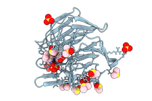
Crystal Structure Of The Keap1 Kelch Domain In Complex With The Xchem Fragment Z19735904 At 1.14 Angstrom Resolution.
Organism: Mus musculus
Method: X-RAY DIFFRACTION Resolution:1.14 Å Release Date: 2025-09-03 Classification: PEPTIDE BINDING PROTEIN Ligands: B0A, SO4, DMS |
 |
Crystal Structure Of The Keap1 Kelch Domain In Complex With The Small Molecule Ucab#827 At 1.40 Angstrom Resolution
Organism: Mus musculus
Method: X-RAY DIFFRACTION Resolution:1.40 Å Release Date: 2025-09-03 Classification: PEPTIDE BINDING PROTEIN Ligands: A1IX2, CL, SO4, DMS |
 |
Crystal Structure Of The Keap1 Kelch Domain In Complex With The Small Molecule Ucab#909 At 1.61 Angstrom Resolution
Organism: Mus musculus
Method: X-RAY DIFFRACTION Resolution:1.61 Å Release Date: 2025-09-03 Classification: PEPTIDE BINDING PROTEIN Ligands: A1IX3, SO4, DMS, CL |
 |
Crystal Structure Of The Keap1 Kelch Domain In Complex With The Small Molecule Ucab#985 At 1.65 Angstrom Resolution
Organism: Mus musculus
Method: X-RAY DIFFRACTION Resolution:1.65 Å Release Date: 2025-09-03 Classification: PEPTIDE BINDING PROTEIN Ligands: A1IX4, SO4, DMS, CL |