
Search Count: 166
All
Selected
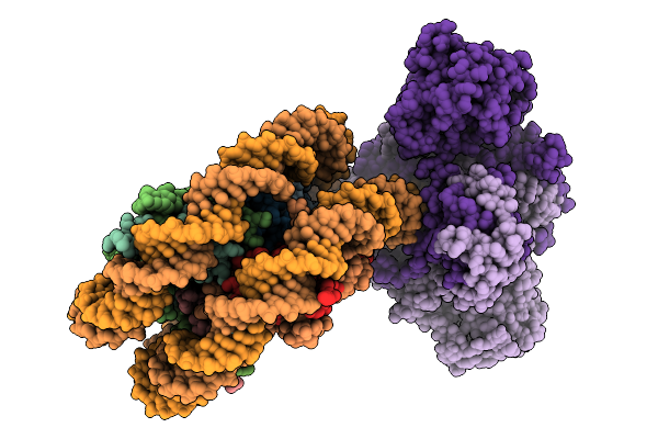 |
Organism: Homo sapiens
Method: ELECTRON MICROSCOPY Resolution:3.32 Å Release Date: 2026-01-14 Classification: DNA BINDING PROTEIN |
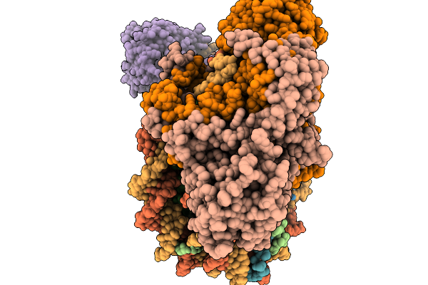 |
Organism: Homo sapiens
Method: ELECTRON MICROSCOPY Resolution:3.56 Å Release Date: 2026-01-14 Classification: DNA BINDING PROTEIN |
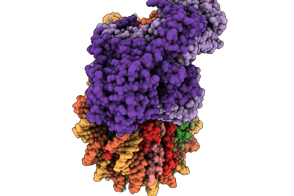 |
Organism: Homo sapiens
Method: ELECTRON MICROSCOPY Resolution:3.39 Å Release Date: 2026-01-14 Classification: DNA BINDING PROTEIN |
 |
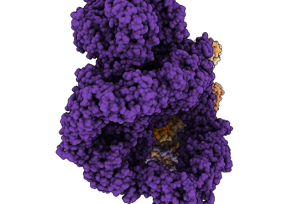
Organism: Homo sapiens
Method: ELECTRON MICROSCOPY Resolution:2.94 Å Release Date: 2026-01-14 Classification: DNA BINDING PROTEIN |
 |
Organism: Homo sapiens
Method: ELECTRON MICROSCOPY Resolution:4.54 Å Release Date: 2026-01-14 Classification: DNA BINDING PROTEIN |
 |
Organism: Homo sapiens
Method: ELECTRON MICROSCOPY Resolution:4.37 Å Release Date: 2026-01-14 Classification: DNA BINDING PROTEIN |
 |
Organism: Homo sapiens
Method: ELECTRON MICROSCOPY Resolution:4.38 Å Release Date: 2026-01-14 Classification: DNA BINDING PROTEIN |
 |
Organism: Homo sapiens
Method: ELECTRON MICROSCOPY Release Date: 2026-01-14 Classification: DNA BINDING PROTEIN |
 |
Organism: Shigella phage sf11 smd-2017
Method: ELECTRON MICROSCOPY Release Date: 2025-11-12 Classification: VIRUS |
 |
Organism: Shigella phage sf11 smd-2017
Method: ELECTRON MICROSCOPY Release Date: 2025-11-12 Classification: VIRUS |
 |
Organism: Shigella phage sf11 smd-2017
Method: ELECTRON MICROSCOPY Resolution:2.40 Å Release Date: 2025-11-12 Classification: VIRUS |
 |
Organism: Bacillus subtilis subsp. subtilis str. 168
Method: ELECTRON MICROSCOPY Resolution:3.40 Å Release Date: 2025-10-15 Classification: MEMBRANE PROTEIN |
 |
Organism: Bacillus subtilis subsp. subtilis str. 168
Method: ELECTRON MICROSCOPY Resolution:2.50 Å Release Date: 2025-10-15 Classification: MEMBRANE PROTEIN Ligands: ADP, VO4, MG, UNL |
 |
Structure Of Lara-Like Nickel-Pincer Nucleotide Cofactor-Utilizing Enzyme With A Single Catalytic Histidine Residue From Streptococcus Plurextorum
Organism: Streptococcus plurextorum
Method: ELECTRON MICROSCOPY Release Date: 2025-09-24 Classification: ISOMERASE Ligands: NI, 4EY |
 |
Structures Of Lara-Like Nickel-Pincer Nucleotide Cofactor-Utilizing Enzyme With A Single Catalytic Histidine Residue From Blautia Wexlerae
Organism: Blautia wexlerae
Method: ELECTRON MICROSCOPY Release Date: 2025-09-17 Classification: ISOMERASE |
 |
Organism: Escherichia phage ms2
Method: ELECTRON MICROSCOPY Release Date: 2025-08-06 Classification: VIRUS LIKE PARTICLE |
 |
Organism: Escherichia phage ms2
Method: ELECTRON MICROSCOPY Release Date: 2025-08-06 Classification: VIRUS LIKE PARTICLE |
 |
Organism: Streptococcus pyogenes
Method: X-RAY DIFFRACTION Resolution:1.43 Å Release Date: 2025-06-25 Classification: PEPTIDE BINDING PROTEIN Ligands: TEW, SO4 |
 |
Organism: Shigella phage sf14
Method: ELECTRON MICROSCOPY Release Date: 2025-02-05 Classification: VIRUS |
 |
Organism: Shigella phage sf14
Method: ELECTRON MICROSCOPY Release Date: 2025-02-05 Classification: VIRUS |