

Search Count: 38
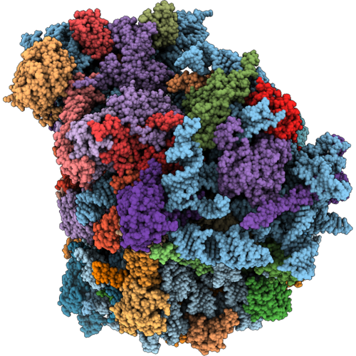 |
Cryoem Structure Of The Vacant Candida Auris 80S Ribosome
Organism: Candidozyma auris
Method: ELECTRON MICROSCOPY Release Date: 2026-09-02 Classification: RIBOSOME Ligands: MG, ZN |
Organism: Candidozyma auris
Method: ELECTRON MICROSCOPY
Release Date: 2026-09-02
Ligands: MG, ZN
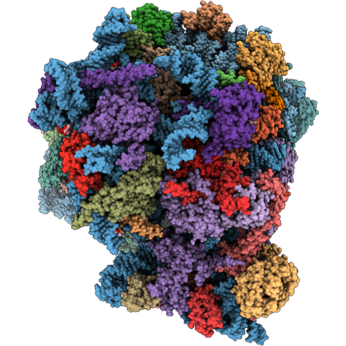 |
Cryoem Structure Of Candida Auris 80S Ribosome In Complex With Cycloheximide And Geneticin G418
Organism: Candidozyma auris
Method: ELECTRON MICROSCOPY Release Date: 2026-09-02 Classification: RIBOSOME Ligands: GET, MG, SPD, 3HE, ZN |
Organism: Candidozyma auris
Method: ELECTRON MICROSCOPY
Release Date: 2026-09-02
Ligands: GET, MG, SPD, 3HE, ZN
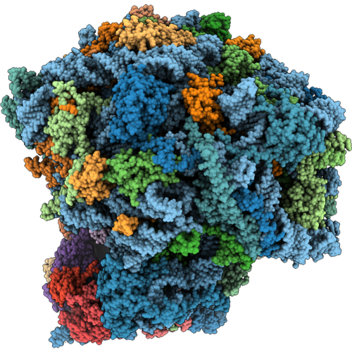 |
Cryoem Structure Of Candida Auris 80S Ribosome In Complex With Blasticidin S
Organism: Candidozyma auris
Method: ELECTRON MICROSCOPY Release Date: 2026-09-02 Classification: RIBOSOME Ligands: MG, BLS, ZN |
Organism: Candidozyma auris
Method: ELECTRON MICROSCOPY
Release Date: 2026-09-02
Ligands: MG, BLS, ZN
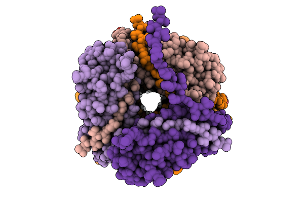 |
Cryo-Em Structure Of The Type I Pilus From Escherichia Coli And The Surrounding Water Network
Organism: Escherichia coli k-12
Method: ELECTRON MICROSCOPY Release Date: 2026-03-11 Classification: CELL ADHESION |
Organism: Escherichia coli k-12
Method: ELECTRON MICROSCOPY
Release Date: 2026-03-11
 |
Cryo-Em Structure Of The Flotillin-Associated Rhodopsin Psfar In Detergent Micelle
Organism: Candidatus pseudothioglobus
Method: ELECTRON MICROSCOPY Resolution:2.56 Å Release Date: 2025-07-23 Classification: MEMBRANE PROTEIN Ligands: LFA |
Organism: Candidatus pseudothioglobus
Method: ELECTRON MICROSCOPY
Release Date: 2025-07-23
Ligands: LFA
 |
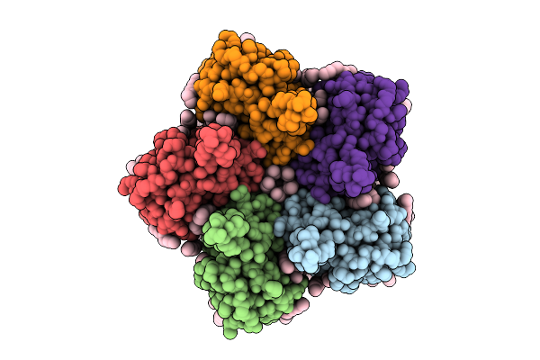
Cryo-Em Structure Of The Light-Driven Proton Pump Pspr In Detergent Micelle
Organism: Candidatus pseudothioglobus sp.
Method: ELECTRON MICROSCOPY Resolution:2.48 Å Release Date: 2025-07-23 Classification: MEMBRANE PROTEIN Ligands: LFA, RET, LMT |
Organism: Candidatus pseudothioglobus sp.
Method: ELECTRON MICROSCOPY
Release Date: 2025-07-23
Ligands: LFA, RET, LMT
 |
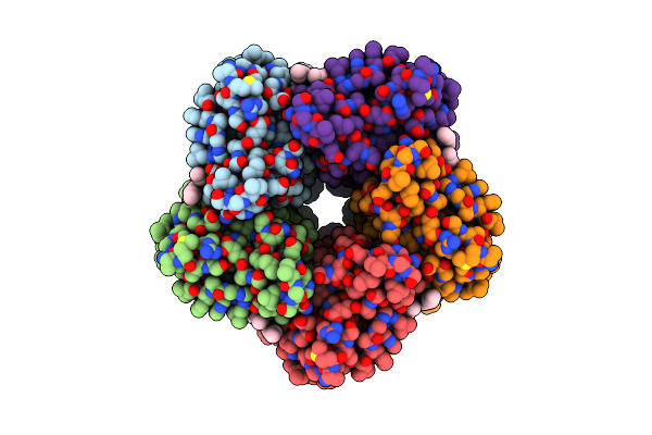
Cryo-Em Structure Of The Double Mutant H84V/E120G Of The Flotillin-Associated Rhodopsin Psfar In Detergent Micelle
Organism: Candidatus pseudothioglobus sp.
Method: ELECTRON MICROSCOPY Resolution:2.81 Å Release Date: 2025-07-23 Classification: MEMBRANE PROTEIN Ligands: LFA, RET |
Organism: Candidatus pseudothioglobus sp.
Method: ELECTRON MICROSCOPY
Release Date: 2025-07-23
Ligands: LFA, RET
 |
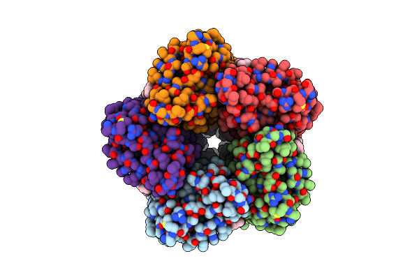
Cryo-Em Structure Of The Microbial Rhodopsin Cryor1 At Ph 4.3 In Detergent
Organism: Cryobacterium levicorallinum
Method: ELECTRON MICROSCOPY Resolution:2.94 Å Release Date: 2025-05-14 Classification: MEMBRANE PROTEIN Ligands: LFA, RET |
Organism: Cryobacterium levicorallinum
Method: ELECTRON MICROSCOPY
Release Date: 2025-05-14
Ligands: LFA, RET
 |
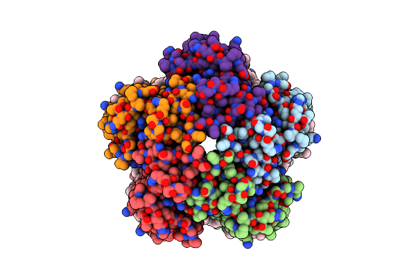
Cryo-Em Structure Of The Microbial Rhodopsin Cryor1 At Ph 8.0 In Nanodisc
Organism: Cryobacterium levicorallinum
Method: ELECTRON MICROSCOPY Resolution:2.43 Å Release Date: 2025-05-14 Classification: MEMBRANE PROTEIN Ligands: LFA, RET |
Organism: Cryobacterium levicorallinum
Method: ELECTRON MICROSCOPY
Release Date: 2025-05-14
Ligands: LFA, RET
 |
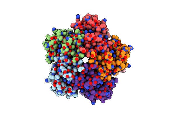
Cryo-Em Structure Of The Microbial Rhodopsin Cryor1 At Ph 8.0 In Detergent
Organism: Cryobacterium levicorallinum
Method: ELECTRON MICROSCOPY Resolution:2.87 Å Release Date: 2025-05-14 Classification: MEMBRANE PROTEIN Ligands: LMT, LFA, RET |
Organism: Cryobacterium levicorallinum
Method: ELECTRON MICROSCOPY
Release Date: 2025-05-14
Ligands: LMT, LFA, RET
 |
Cryo-Em Structure Of The Microbial Rhodopsin Cryor1 At Ph 10.5 In Detergent In The Ground State
Organism: Cryobacterium levicorallinum
Method: ELECTRON MICROSCOPY Resolution:2.70 Å Release Date: 2025-05-14 Classification: MEMBRANE PROTEIN Ligands: LMT, RET, LFA |
Organism: Cryobacterium levicorallinum
Method: ELECTRON MICROSCOPY
Release Date: 2025-05-14
Ligands: LMT, RET, LFA
 |
Cryo-Em Structure Of The Microbial Rhodopsin Cryor1 At Ph 10.5 In Detergent In The M State
Organism: Cryobacterium levicorallinum
Method: ELECTRON MICROSCOPY Resolution:2.30 Å Release Date: 2025-05-14 Classification: MEMBRANE PROTEIN Ligands: LMT, RET |
Organism: Cryobacterium levicorallinum
Method: ELECTRON MICROSCOPY
Release Date: 2025-05-14
Ligands: LMT, RET
 |
Cryo-Em Structure Of The Microbial Rhodopsin Cryor2 At Ph 8.0 In Detergent
Organism: Subtercola endophyticus
Method: ELECTRON MICROSCOPY Resolution:2.44 Å Release Date: 2025-05-14 Classification: MEMBRANE PROTEIN Ligands: LFA, RET |
Organism: Subtercola endophyticus
Method: ELECTRON MICROSCOPY
Release Date: 2025-05-14
Ligands: LFA, RET
 |
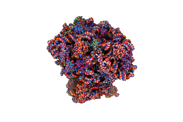
Structure Of Candida Albicans 80S Ribosome In Complex With Mefloquine (Non-Rotated State)
Organism: Candida albicans sc5314
Method: ELECTRON MICROSCOPY Release Date: 2025-04-23 Classification: RIBOSOME Ligands: ZN, YMZ, SPK |
Organism: Candida albicans sc5314
Method: ELECTRON MICROSCOPY
Release Date: 2025-04-23
Ligands: ZN, YMZ, SPK
 |
The Structure Of The Candida Albicans Ribosome With Trna-Fmet, Mrna, And Compounds (Gen And Mfq) Shows Strong Density For The A Site Trna
Organism: Candida albicans
Method: ELECTRON MICROSCOPY Release Date: 2025-04-23 Classification: RIBOSOME Ligands: SPK, GET, ZN |
Organism: Candida albicans
Method: ELECTRON MICROSCOPY
Release Date: 2025-04-23
Ligands: SPK, GET, ZN
 |
The Structure Of The Candida Albicans Ribosome With Trna-Fmet, Mrna, And Compounds (Gen And Mfq) With Strong Density For The P-Site Trna
Organism: Escherichia coli, Candida albicans sc5314
Method: ELECTRON MICROSCOPY Release Date: 2025-04-23 Classification: RIBOSOME Ligands: SPK, GET, ZN, YMZ |
Organism: Escherichia coli, Candida albicans sc5314
Method: ELECTRON MICROSCOPY
Release Date: 2025-04-23
Ligands: SPK, GET, ZN, YMZ
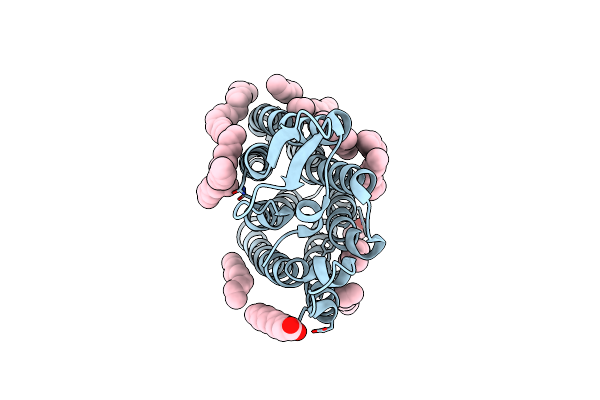 |
Crystal Structure Of The Light-Driven Sodium Pump Ernar In The Monomeric Form At Ph 4.6
Organism: Erythrobacter
Method: X-RAY DIFFRACTION Resolution:1.70 Å Release Date: 2024-04-24 Classification: MEMBRANE PROTEIN Ligands: LFA, OLA |
Organism: Erythrobacter
Method: X-RAY DIFFRACTION
Release Date: 2024-04-24
Ligands: LFA, OLA
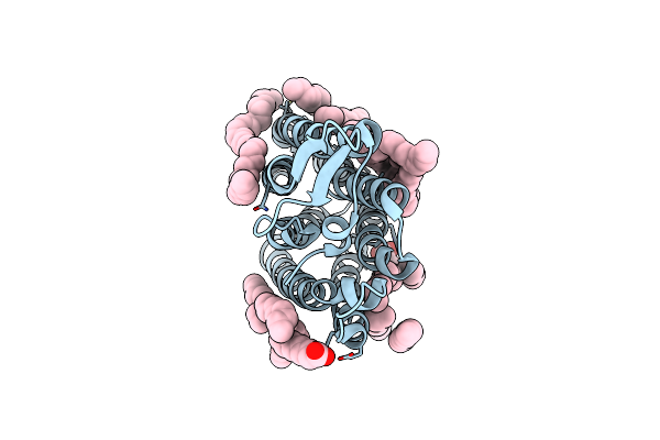 |
Crystal Structure Of The Light-Driven Sodium Pump Ernar In The Monomeric Form At Ph 8.8
Organism: Erythrobacter
Method: X-RAY DIFFRACTION Resolution:1.71 Å Release Date: 2024-04-24 Classification: MEMBRANE PROTEIN Ligands: LFA, OLA |
Organism: Erythrobacter
Method: X-RAY DIFFRACTION
Release Date: 2024-04-24
Ligands: LFA, OLA
 |
Cryo-Em Structure Of The Light-Driven Sodium Pump Ernar In The Pentameric Form At Ph 8.0
Organism: Erythrobacter
Method: ELECTRON MICROSCOPY Resolution:2.63 Å Release Date: 2024-04-24 Classification: MEMBRANE PROTEIN Ligands: LFA, LMT |
Organism: Erythrobacter
Method: ELECTRON MICROSCOPY
Release Date: 2024-04-24
Ligands: LFA, LMT
 |
Cryo-Em Structure Of The Light-Driven Sodium Pump Ernar In The Pentameric Form At Ph 4.3
Organism: Erythrobacter
Method: ELECTRON MICROSCOPY Resolution:2.50 Å Release Date: 2024-04-24 Classification: MEMBRANE PROTEIN Ligands: LMT, LFA |
Organism: Erythrobacter
Method: ELECTRON MICROSCOPY
Release Date: 2024-04-24
Ligands: LMT, LFA