

Search Count: 21
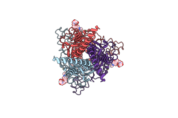 |
Cryo-Em Structure Of P2X3 Receptor In Complex With Camlipixant
Organism: Canis lupus
Method: ELECTRON MICROSCOPY Release Date: 2025-03-19 Classification: MEMBRANE PROTEIN Ligands: A1AQX, NAG |
Organism: Canis lupus
Method: ELECTRON MICROSCOPY
Release Date: 2025-03-19
Ligands: A1AQX, NAG
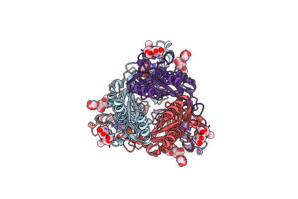 |
Cryo-Em Structure Of P2X3 Receptor In Complex With Atp:Mg2+
Organism: Canis lupus
Method: ELECTRON MICROSCOPY Release Date: 2025-03-19 Classification: MEMBRANE PROTEIN Ligands: NAG, MG, ATP |
Organism: Canis lupus
Method: ELECTRON MICROSCOPY
Release Date: 2025-03-19
Ligands: NAG, MG, ATP
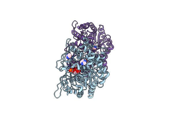 |
The Structure Of Gan1D W433A In Complex With Galactose-6P
Organism: Geobacillus stearothermophilus
Method: X-RAY DIFFRACTION Resolution:1.78 Å Release Date: 2022-10-19 Classification: HYDROLASE Ligands: IMD, BGP, GOL |
Organism: Geobacillus stearothermophilus
Method: X-RAY DIFFRACTION
Release Date: 2022-10-19
Ligands: IMD, BGP, GOL
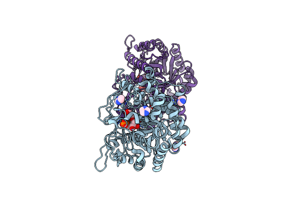 |
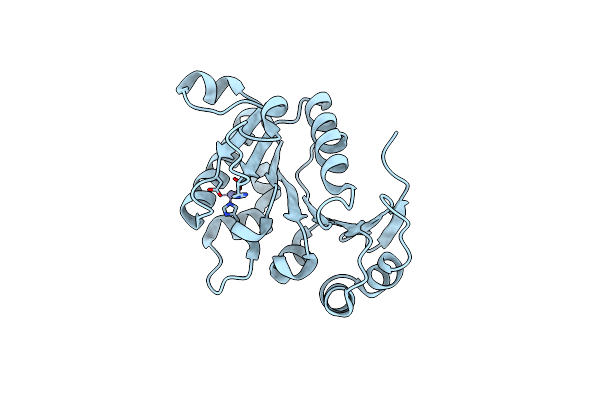
The Structure Of Gan1D W433A In Complex With Cellobiose-6-Phosphate
Organism: Geobacillus stearothermophilus
Method: X-RAY DIFFRACTION Resolution:1.59 Å Release Date: 2022-10-19 Classification: HYDROLASE Ligands: IMD |
Organism: Geobacillus stearothermophilus
Method: X-RAY DIFFRACTION
Release Date: 2022-10-19
Ligands: IMD
 |
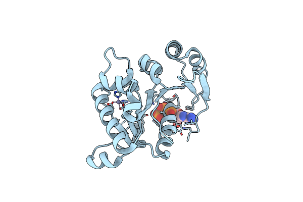
The Crystal Structure Of B204, The Dna-Packaging Atpase From Sulfolobus Turreted Icosahedral Virus
Organism: Sulfolobus turreted icosahedral virus 1
Method: X-RAY DIFFRACTION Resolution:1.96 Å Release Date: 2016-01-13 Classification: VIRAL PROTEIN Ligands: ZN |
Organism: Sulfolobus turreted icosahedral virus 1
Method: X-RAY DIFFRACTION
Release Date: 2016-01-13
Ligands: ZN
 |
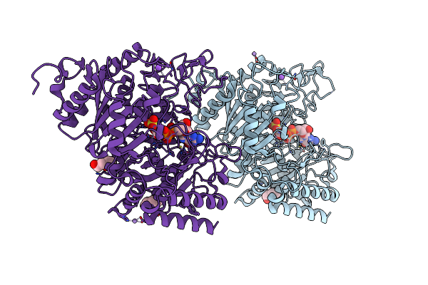
The Crystal Structure Of Stiv B204 Complexed With Amp-Pnp
Organism: Sulfolobus turreted icosahedral virus 1
Method: X-RAY DIFFRACTION Resolution:2.05 Å Release Date: 2016-01-13 Classification: VIRAL PROTEIN Ligands: ZN, ANP |
Organism: Sulfolobus turreted icosahedral virus 1
Method: X-RAY DIFFRACTION
Release Date: 2016-01-13
Ligands: ZN, ANP
 |
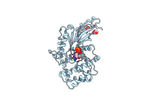
I45T Cytosolic Phosphoenolpyruvate Carboxykinase In Complex With Beta-Sulfopyruvate And Gtp
Organism: Rattus norvegicus
Method: X-RAY DIFFRACTION Resolution:2.00 Å Release Date: 2014-06-11 Classification: LYASE Ligands: MN, GTP, SPV, NA, 1PE, ETX |
Organism: Rattus norvegicus
Method: X-RAY DIFFRACTION
Release Date: 2014-06-11
Ligands: MN, GTP, SPV, NA, 1PE, ETX
 |
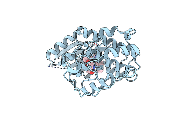
Crystal Structure Of The G202D Mutant Of Human G-Alpha-I1
Organism: Homo sapiens
Method: X-RAY DIFFRACTION Resolution:2.04 Å Release Date: 2012-10-24 Classification: SIGNALING PROTEIN Ligands: GDP, SO4, CL, GOL |
Organism: Homo sapiens
Method: X-RAY DIFFRACTION
Release Date: 2012-10-24
Ligands: GDP, SO4, CL, GOL
 |
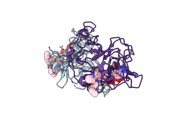
Crystal Structure Of Cdk2 In Complex With Inhibitor Bs-194
Organism: Homo sapiens
Method: X-RAY DIFFRACTION Resolution:1.78 Å Release Date: 2010-12-08 Classification: TRANSFERASE/TRANSFERASE INHIBITOR Ligands: NS9 |
Organism: Homo sapiens
Method: X-RAY DIFFRACTION
Release Date: 2010-12-08
Ligands: NS9
 |
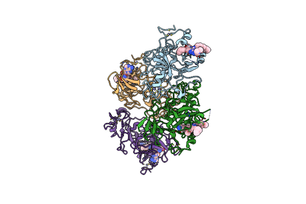
Human G9A-Like (Glp, Also Known As Ehmt1) In Complex With Inhibitor E11
Organism: Homo sapiens
Method: X-RAY DIFFRACTION Resolution:2.78 Å Release Date: 2010-06-30 Classification: TRANSFERASE Ligands: SAH, E11, ZN, EDO |
Organism: Homo sapiens
Method: X-RAY DIFFRACTION
Release Date: 2010-06-30
Ligands: SAH, E11, ZN, EDO
 |
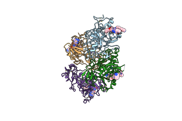
Human G9A-Like (Glp, Also Known As Ehmt1) In Complex With Inhibitor E67
Organism: Homo sapiens
Method: X-RAY DIFFRACTION Resolution:2.49 Å Release Date: 2010-06-30 Classification: TRANSFERASE Ligands: ZN, E67, SAH |
Organism: Homo sapiens
Method: X-RAY DIFFRACTION
Release Date: 2010-06-30
Ligands: ZN, E67, SAH
 |
Human G9A-Like (Glp, Also Known As Ehmt1) In Complex With Inhibitor E72
Organism: Homo sapiens
Method: X-RAY DIFFRACTION Resolution:2.14 Å Release Date: 2010-06-30 Classification: TRANSFERASE Ligands: ZN, E72, SAH |
Organism: Homo sapiens
Method: X-RAY DIFFRACTION
Release Date: 2010-06-30
Ligands: ZN, E72, SAH
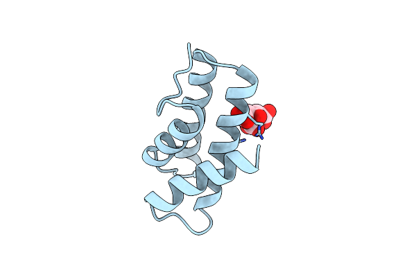 |
Crystal Structure Of Yellowstone Sirv Coat Protein C-Terminus
Organism: Sulfolobus islandicus rudivirus 1 variant ynp
Method: X-RAY DIFFRACTION Resolution:1.67 Å Release Date: 2009-04-21 Classification: VIRAL PROTEIN Ligands: CIT |
Organism: Sulfolobus islandicus rudivirus 1 variant ynp
Method: X-RAY DIFFRACTION
Release Date: 2009-04-21
Ligands: CIT
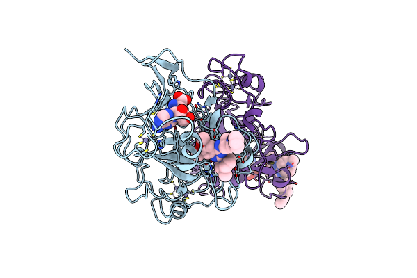 |
G9A-Like Protein Lysine Methyltransferase Inhibition By Bix-01294
Organism: Homo sapiens
Method: X-RAY DIFFRACTION Resolution:2.40 Å Release Date: 2009-02-17 Classification: TRANSFERASE Ligands: SAH, Q4A, ZN |
Organism: Homo sapiens
Method: X-RAY DIFFRACTION
Release Date: 2009-02-17
Ligands: SAH, Q4A, ZN
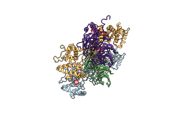 |
The Multifunctional Nature Of Gbeta5/Rgs9 Revealed From Its Crystal Structure
Organism: Mus musculus
Method: X-RAY DIFFRACTION Resolution:1.95 Å Release Date: 2008-01-29 Classification: SIGNALING PROTEIN Ligands: GOL |
Organism: Mus musculus
Method: X-RAY DIFFRACTION
Release Date: 2008-01-29
Ligands: GOL
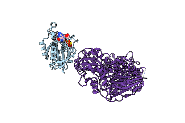 |
Activated Rac1 Bound To Its Effector Phospholipase C Beta 2
Organism: Homo sapiens
Method: X-RAY DIFFRACTION Resolution:2.20 Å Release Date: 2006-11-21 Classification: SIGNALING PROTEIN,APOPTOSIS/HYDROLASE Ligands: MG, GSP, CA |
Organism: Homo sapiens
Method: X-RAY DIFFRACTION
Release Date: 2006-11-21
Ligands: MG, GSP, CA
 |
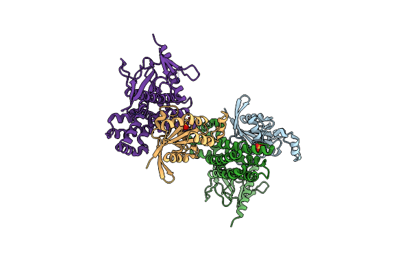
The Binding Mode Of Epothilone A On A,B-Tubulin By Electron Crystallography
Organism: Bos taurus
Method: ELECTRON CRYSTALLOGRAPHY Resolution:2.89 Å Release Date: 2004-09-14 Classification: CELL CYCLE, STRUCTURAL PROTEIN Ligands: GTP, GDP, EP |
Organism: Bos taurus
Method: ELECTRON CRYSTALLOGRAPHY
Release Date: 2004-09-14
Ligands: GTP, GDP, EP
 |
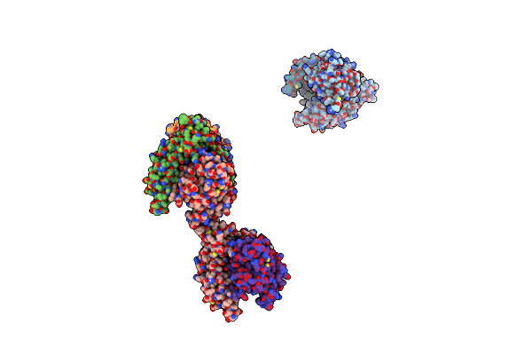
Guanine Nucleotide Exchange Region Of Intersectin In Complex With Cdc42
Organism: Homo sapiens
Method: X-RAY DIFFRACTION Resolution:2.30 Å Release Date: 2002-05-29 Classification: SIGNALING PROTEIN Ligands: SO4 |
Organism: Homo sapiens
Method: X-RAY DIFFRACTION
Release Date: 2002-05-29
Ligands: SO4
 |
Crystal Structure Of The Dbl And Pleckstrin Homology Domains Of Dbs In Complex With Rhoa
Organism: Mus musculus, Homo sapiens
Method: X-RAY DIFFRACTION Resolution:2.81 Å Release Date: 2002-05-29 Classification: SIGNALING PROTEIN |
Organism: Mus musculus, Homo sapiens
Method: X-RAY DIFFRACTION
Release Date: 2002-05-29
 |
Crystal Structure Of The Dh/Ph Fragment Of Murine Dbs In Complex With The Placental Isoform Of Human Cdc42
Organism: Mus musculus, Homo sapiens
Method: X-RAY DIFFRACTION Resolution:2.40 Å Release Date: 2002-03-20 Classification: SIGNALING PROTEIN |
Organism: Mus musculus, Homo sapiens
Method: X-RAY DIFFRACTION
Release Date: 2002-03-20